
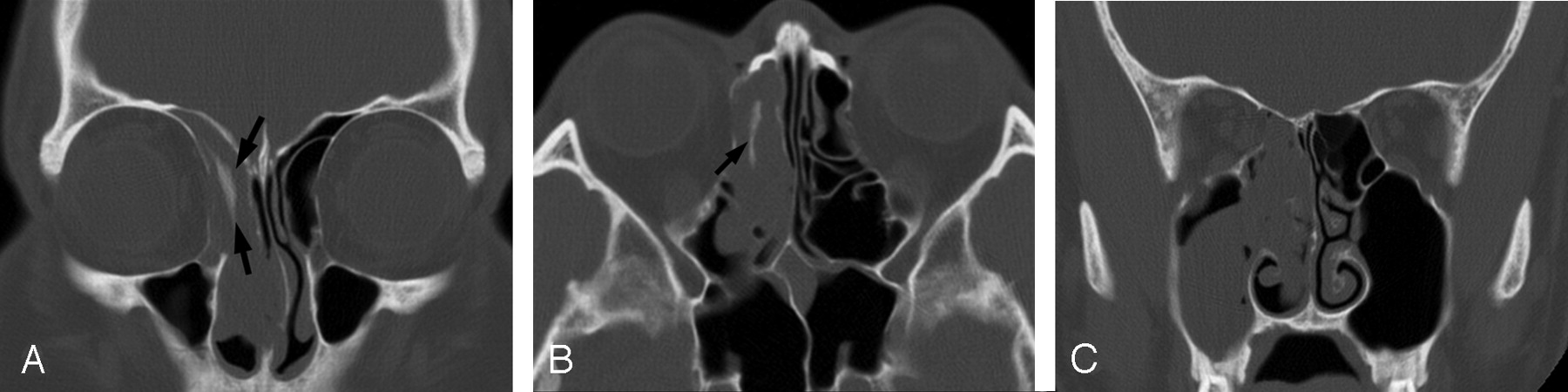
- Fig 1.
CT images of a patient with inverted papilloma.
A and B, Axial and coronal CT images show focal plaquelike hyperostosis in part of right ethmoid sinuses (arrows).
C, Although tumor extends to the right maxillary sinus and nasal cavity, no additional foci of hyperostosis are seen. Intraoperative endoscopic examination confirmed the limitation of tumor origin to the ethmoid sinuses.
- Fig 2.
CT and MR images of patients with inverted papilloma of the maxillary sinus.
A, Axial CT image of a patient with inverted papilloma shows cone-shaped focal hyperostosis involving the posterior wall of the left maxillary sinus (arrows).
B, Sagittal T2-weighted MR image of the patient clearly shows the centrifugal pattern of tumor growth with a hyperostotic focus (white arrow) at the posterior wall of the left maxillary sinus, which was confirmed to be a tumor origin by surgery.
C, Axial CT image of another patient shows cone-shaped hyperostosis (arrow) involving the anterior wall of the left maxillary sinus, which was proved to be the origin of inverted papilloma by intraoperative endoscopy.
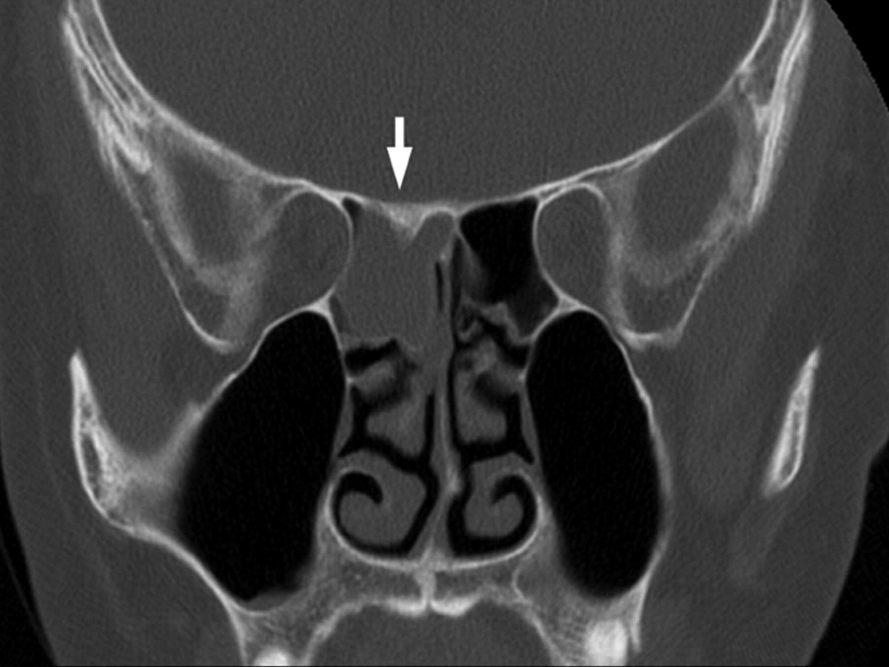
- Fig 3.
Coronal CT image in a patient with inverted papilloma shows localized cone-shaped hyperostosis of the superior wall of the posterior ethmoid sinus (white arrow). Intraoperative endoscopic examination confirmed that the origin of tumor was located at the superior wall of the right posterior ethmoid sinus.


- Copyright © American Society of Neuroradiology


