fig 4.

fig 4.
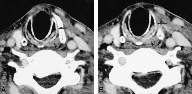
A and B, Two consecutive axial CT scans in a patient with a right-sided glomus vagale show atrophy of the right PCA muscle and right cricothyroid muscle relative to the normal left side (arrowhead indicates normal left PCA muscle; arrow, normal left cricothyroid muscle)


