fig 3.

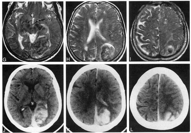
Case 2: 79-year-old woman with acute IVH, intraparenchymal hemorrhage, and SAH who had imaging on the day of ictus. MR and CT studies were obtained 2 hours apart. The FLAIR images show the IVH and SAH better than the T1- and T2-weighted images or the CT scans do.
A–C, FLAIR images (6700/150/2, TI = 2200) show large left parietooccipital intraparenchymal hemorrhage with intraventricular extension and associated SAH. The intraparenchymal hemorrhage is of mixed signal (mostly isointense). However, the IVH is well seen as hyperintense in the right occipital horn (arrow, A) and left body (arrow, B) of the lateral ventricles. SAH is well seen as hyperintensities in the right dorsal frontoparietal sulci (B and C).
D–I, T1-weighted (450/20/2) (D–F) and T2-weighted (2000/120/2) (G–I) images do not show the IVH and SAH as conspicuously as the FLAIR images do.
J–L, Noncontrast CT scans readily show the hyperdense intraparenchymal hemorrhage. However, the IVH and SAH are shown more conspicuously on the FLAIR images.


Related Articles
Cited By...
- Correlation of clot imaging with endovascular recanalization in internal carotid artery terminus occlusion
- Double Inversion Recovery MR Sequence for the Detection of Subacute Subarachnoid Hemorrhage
- Characterization of Arterial Thrombus Composition by Magnetic Resonance Imaging in a Swine Stroke Model
- 3D Fluid-Attenuated Inversion Recovery Imaging: Reduced CSF Artifacts and Enhanced Sensitivity and Specificity for Subarachnoid Hemorrhage
- Exclusion of brain lesions: is MR contrast medium required after a negative fluid-attenuated inversion recovery sequence?
- Current theory in imaging of intracranial vascular disease
- Diffusion-weighted MRI as an evolving standard of care in acute stroke
- Is fluid-attenuated inversion recovery MRI more sensitive than conventional MRI for hypoglycemic brain injury?
- Intraventricular CSF Pulsation Artifact on Fast Fluid-Attenuated Inversion-Recovery MR Images: Analysis of 100 Consecutive Normal Studies