fig 4.

fig 4.
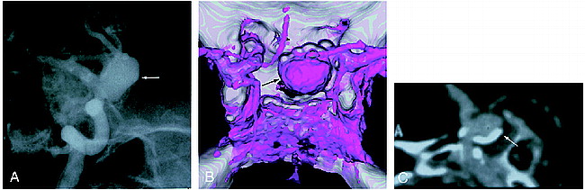
;t1Case 13: Patient with an aneurysm of the internal carotid artery–posterior communicating artery.
A, Right carotid angiogram (left anterior oblique view) shows an aneurysm (arrow).
B, 3D CT angiogram with SSD (superior view) shows an aneurysm (arrow). Mural calcium cannot be well appreciated.
C, Composite sagittal CPR image clearly shows calcification in the wall of the aneurysm (arrow) and parent artery.


