fig 2.

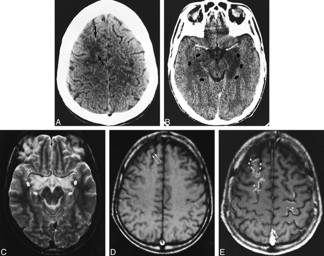
Case 2: 28-year-old man who underwent bone marrow transplantation and immunosuppressive therapy with CsA.
A and B, Axial CT scans obtained at the onset of right arm paresis, mutism, and mydriatic pupils show confluent hypodensity in the right frontal white matter (black arrows) and patchy areas of hypodensity in the left prerolandic area (white arrows) (A). After contrast injection (B), there is marked hypodensity of the left hippocampus and slight hypodensity in the right hippocampus (arrows).
C–E, MR studies 20 days later. Axial T2-weighted image at the level of the midbrain (C) shows partial resolution of the left hippocampal lesion with remaining bilateral hippocampal hyperintensities (arrows). Axial noncontrast T1-weighted image in the frontoparietal region (D) shows a subtle hyperintensity in the right frontal cortex (arrows). T1-weighted image after contrast administration (E) shows gyral enhancement in both frontal and parietal lobes (arrowheads).


