fig 3.

fig 3.
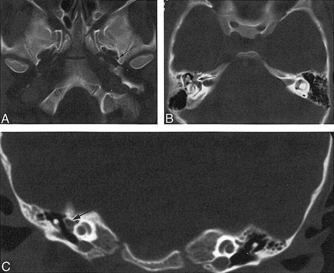
Case 3: 6-year-old girl with vertigo.
A, Axial CT scan shows a normal left foramen spinosum (arrow) and absence of the right foramen spinosum.
B, Axial CT scan through middle ear shows prominent soft tissue, representing facial nerve and PSA (arrow).
C, Coronal CT scan through middle ear shows prominent soft tissue, representing facial nerve and PSA (arrow).


Related Articles
Cited By...
- Cerebral neurovascular embryology, anatomic variations, and congenital brain arteriovenous lesions
- The Many Faces of Persistent Stapedial Artery: CT Findings and Embryologic Explanations
- Middle Meningeal Artery: Anatomy and Variations
- Stapedial Artery: From Embryology to Different Possible Adult Configurations
- Temporal Bone CT: Anatomy, Technique, and Associated Pathophysiology
- A Venous Cause for Facial Canal Enlargement: Multidetector Row CT Findings and Histopathologic Correlation
- Dangerous Extracranial-Intracranial Anastomoses and Supply to the Cranial Nerves: Vessels the Neurointerventionalist Needs to Know
- Persistent Stapedial Artery: MR Angiographic and CT Findings
- Bilateral Aberrant Internal Carotid Arteries with Bilateral Persistent Stapedial Arteries and Bilateral Duplicated Internal Carotid Arteries