
Abstract
Summary: The case reports of 17-year-old female dizygotic twins are presented. One of the twins had bilateral closed-lip schizencephaly, and the other had focal cortical dysplasia. Septum pellucidum was absent in both cases. The cortical dysplasia in case 2 corresponded to the same hemispheric location with the right schizencephalic cleft in case 1. The combination of schizencephaly and focal cortical dysplasia in siblings or twins has not been previously reported.
Schizencephaly has been proposed as an extreme variation of focal cortical dysplasia (1). Although one hypothesis for its cause is based on vascular compromise during early neuroembryogenesis (2), familial occurrence also has been reported (3–5). However, the ethiopathogenesis of schizencephaly is still unclear.
Case Reports
Case 1
A 17-year-old right-handed female patient was referred for a cranial MR imaging examination. She had intractable epilepsy characterized by simple partial, complex partial, generalized, and secondary generalized seizures since the age of7 years. She had mild mental retardation, although her motor development was normal. There was nothing remarkable in her prenatal and natal history. A neurologic examination did not reveal any additional findings. The results of an ophthalmologic examination were normal, and the patient's hormonal development was normal. Growth hormone and adrenocorticotrophic hormone levels were within normal ranges. On cranial MR images, bilateral parietal clefts lined by gray matter were detected. The clefts were typical of closed-lip schizencephaly, showing no communication of the lateral ventricles and subarachnoid space. Septum pellucidum was absent (Fig 1). Optic nerves and chiasm were normal. The interhemispheric fissure was intact.

Image from the case of a 17-year-old female patient (case 1). T2-weighted axial image shows bilateral closed-lip schizencephaly and absence of the septum pellucidum.
Case 2
The dizygotic twin of case 1 was also referred for cranial MR imaging to investigate the cause of her generalized convulsions, which had been occurring for 1 year. She was also right-handed and phenotypically different from her twin sister. She had been taking antiepileptics for 1 year, although irregularly, and she was not receiving antiepileptic drugs at the time of referral. She had a better mental and neurodevelopmental status compared with the patient in case 1. The results of her physical, neurologic, and ophthalmologic examinations were normal. On cranial MR images, a focal cortical dysplasia was noted, which was in the same hemispheric location as the schizencephalic cleft in case 1 (Fig 2). Septum pellucidum was absent. Optic nerves and chiasm were normal, and the interhemispheric fissure was intact. The parents and two older siblings of the twins did not have a neurologic disease. The results of their neurologic and cranial MR examinations were normal.
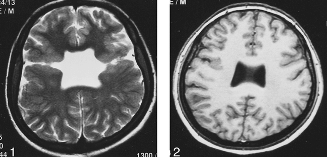
Image from the case of a 17-year-old female patient (case 2). 3D fast field–echo axial image shows septum pellucidum agenesis and right focal cortical dysplasia
Discussion
Schizencephaly is a rare developmental disorder, defined as an abnormal gray matter–lined cleft extending from the pial surface to the ventricle. Its spectrum ranges from a narrow cleavage (closed-lip schizencephaly) to a wide communication between the subarachnoid space and the ventricle (open-lip schizencephaly) (6). Focal cortical dysplasia is among the most commonly associated anomalies with schizencephaly. Evidence from anatomic and clinical studies suggests that schizencephaly and focal cortical dysplasia are the result of the same pathologic process (1). According to Barkovich and Kjos (1), if this process involves only the superficial layer of the fetal brain, a disorganized and thickened cortex, focal cortical dysplasia, develops. If the entire thickness of the cerebral hemisphere is affected, the result is a cleft connecting the subarachnoid space and the lateral ventricle (1).
Toxic exposure, viral infections, genetic factors, and vascular, familial, or metabolic involvement have been suggested to explain the complex cause of schizencephaly (6, 7). There have been few reports indicating familial occurrence in which schizencephaly was detected in siblings (3–8). Twins with schizencephaly have been reported in one study (6). In our cases, one of the twins had bilateral closed-lip schizencephaly and the other had focal cortical dysplasia, both of which were located around the sylvian fissure. To our knowledge, such a combination has not been reported in siblings. Focal cortical dysplasia in case 2 may be considered a form of schizencephaly in which there is an incomplete cleft that does not extend down to the ventricular margin. It was also noteworthy that the location of focal cortical dysplasia in one twin corresponded exactly to that of the schizencephalic cleft in the right parietal lobe in the other. Barkovich and Kjos (1) and Packard et al (6) noted that schizencephaly and focal cortical dysplasia often occur in similar hemispheric locations; however, this has not been reported to occur separately in dizygotic twins.
Agenesis of the septum pellucidum is a rare disorder and, when identified, is almost always associated with some intracranial anomalies, including schizencephaly (9). The septum pellucidum is reported to be absent in approximately 80% to 90% of the schizencephalic patients (6, 10). The septum pellucidum was absent in both of our cases. In case 2, septum pellucidum agenesis is accompanied by focal cortical dysplasia. Focal cortical dysplasia has not been reported among the disorders accompanying septum pellucidum agenesis (9, 11). However, focal cortical dysplasia, at least in some forms, may be considered to be a minor variation of schizencephaly (1).
The clinical manifestations of schizencephaly depend on the extent of the lesion. Unilateral forms of schizencephaly generally present milder symptoms than do bilateral forms. The clinical severity is proportional to the number of involved lobes. Hemiparesis, seizure, and low intellectual level are frequent symptoms (6). Case 1 had more severe clinical presentation as well as lower language skills and intellectual level than did her twin. These differences of clinical presentation also support the idea that focal cortical dysplasia is a minor variation of schizencephaly.
There was nothing remarkable regarding the prenatal histories of our patients. There is a possibility, although we think it is small, that the findings in our cases are due only to coincidence. Although the occurrence of schizencephaly and focal cortical dysplasia in our dizygotic twins could be a result of familial factors, a common prenatal injury could have affected the brains of the fetuses to varying extents. Whatever the reason, it did not cause the same malformations in the twins. Thus, even if a familial occurrence is present, a complex cause of different malformations in the twins is suggested.
In conclusion, our findings support the theory that schizencephaly and focal cortical dysplasia are variations of each other with a common pathogenetic origin. Milder forms of this entity may be present in the twins of the patients with schizencephaly.
Acknowledgments
We thank Teresa Soylu for kind help in editing the manuscript.
Footnotes
↵1 Address reprint requests to Dr. Utku Senol, Department of Radiology, Faculty of Medicine, Akdeniz University, 07070 Antalya, Turkey.
References
- Received January 18, 2000.
- Copyright © American Society of Neuroradiology
In this issue


Jump to section
Related Articles
Cited By...
- No citing articles found.