fig 1.

fig 1.
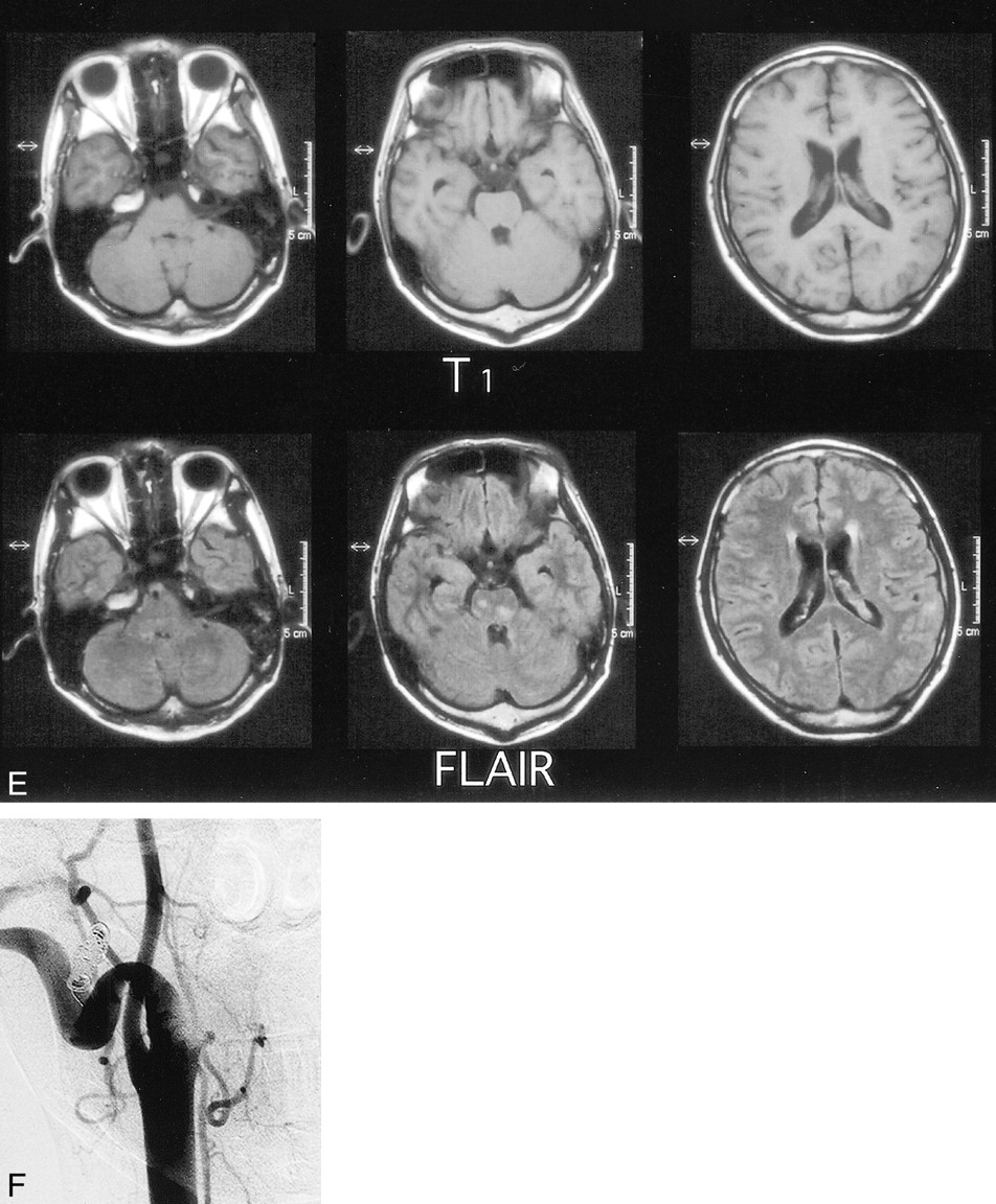
fig. 1 Continued.
E, Axial FLAIR MR images (6000/120) after embolization show only a few areas of high signal intensity in the brain stem and cerebellum.
F, Angiogram 1 month after admission shows that the external carotid artery and jugular vein are almost equivalent in size, and no arteriovenous communication is detected.


Related Articles
Cited By...
- No citing articles found.