fig 5.
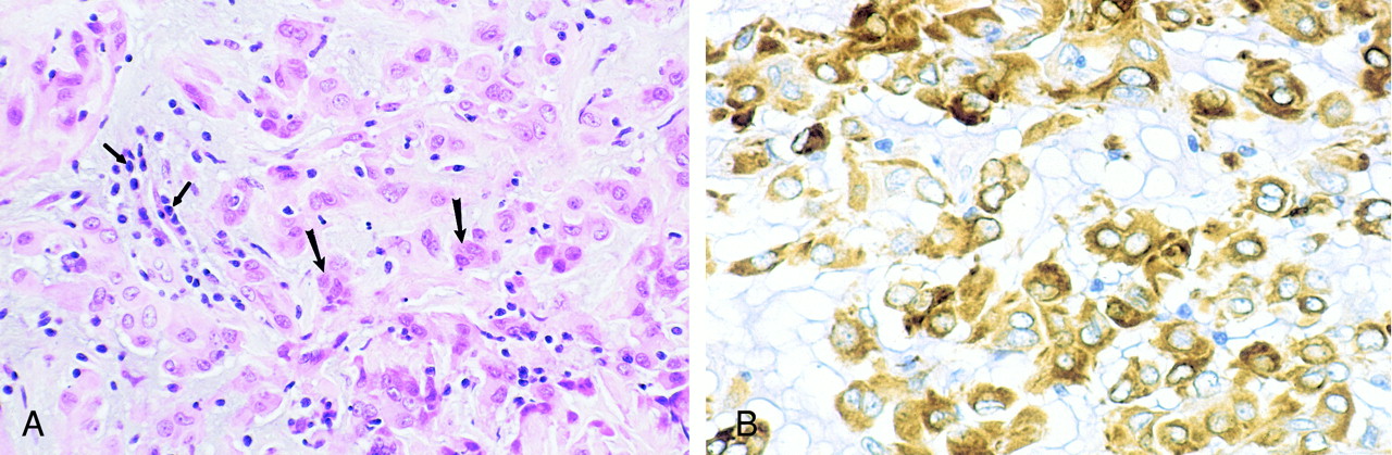
fig 5.
Photomicrographs of chordoid glioma (Patient 5).
A, Hematoxylin and eosin stain of chordoid glioma reveals cords and clusters of eosinophilic epithelioid tumor cells (large arrows) dispersed within a slightly bluish mucinous matrix. A lymphoplasmacytic infiltrate is consistently present (small arrows).
B, Glial fibrillary acidic protein stain of chordoid glioma tumor cells shows strong, diffuse, cytoplasmic immunoreactivity.


