fig 1.
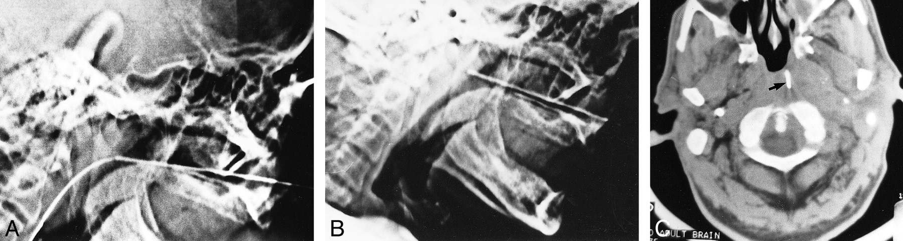
fig 1.
Case 2: 64-year-old man undergoing transnasal access for sampling a skull base lesion.
A and B, Lateral CT scout images. Metal canula was advanced over a guidewire placed transnasally (A). After removal of the guidewire, the canula was directed slightly superiorly to the nasopharyngeal mass (B).
C, Axial CT scan shows the tip of the metal canula in the nasopharyngeal lesion (arrow).


Related Articles
Cited By...
- No citing articles found.