Fig 2.
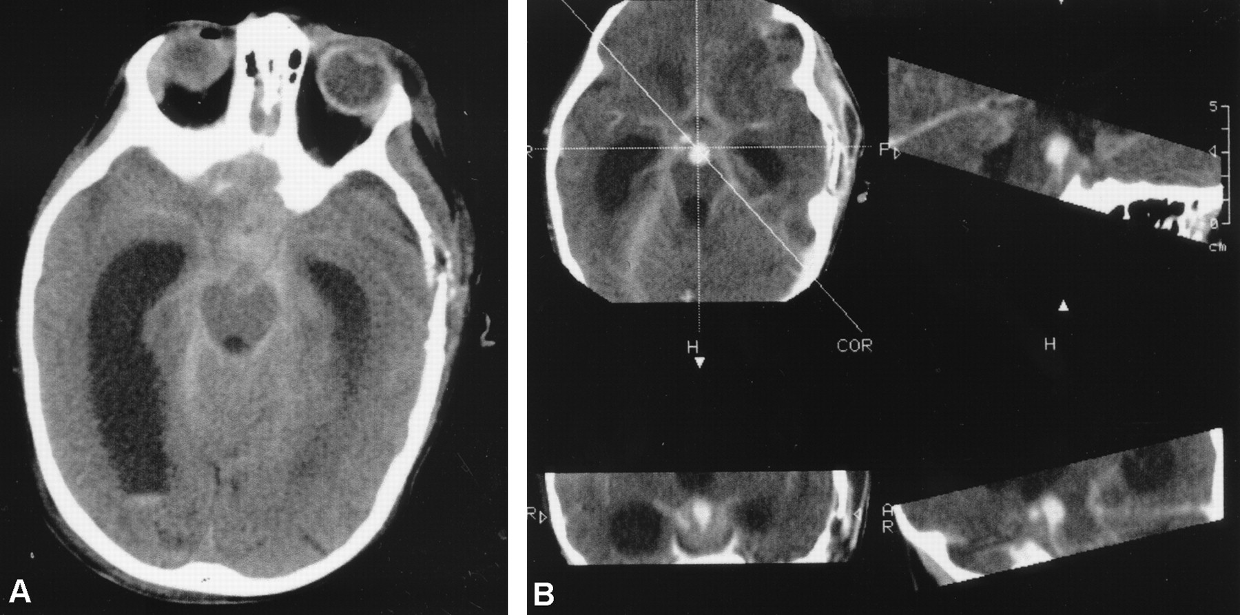
Fig 2.
CT scans of the brain obtained on postoperative day 8.
A, CT scan shows subarachnoid and intraventricular hemorrhage, associated with hydrocephalus. New, irregularly roundish lesions surrounded by the hyperattenuated blood can be seen inside the interpeduncular cistern.
B, Contrast-enhanced CT scans show intense enhancement of these lesions, confirming the hypothesis of aneurysms of the basilar and right posterior cerebral arteries.


