
Abstract
BACKGROUND AND PURPOSE: Few studies have concerned the absolute apparent diffusion coefficient (ADC) values in the normal human brain and the effect of aging on diffusion. Therefore, our purpose was to determine whether the average ADC (ADCav) values in the various regions of the brain differ with age, sex, or hemisphere and to establish reference values of the absolute ADCav for further studies.
METHODS: Subjects (40 men and 40 women) were chosen from a healthy population; age groups were 20–34, 35–49, and 50–64 years and 65 years or older (n = 20 each). All subjects were examined with MR imaging, including conventional and diffusion-weighted imaging in three orthogonal directions with two b values (0 and 1000 s/mm2) at 1.5 T. Bilateral ADCav values were determined in 36 regions of interest encompassing the entire brain.
RESULTS: ADCav values were highest in the cortical gray matter ([0.89 ± 0.04] × 10−3 mm2/s; range, 0.78–1.09 × 10⁻³), lower in the deep gray matter ([0.75 ± 0.03] × 10−3 mm2/s; range, 0.64–0.83 × 10⁻³), and lowest in the white matter ([0.70 ± 0.03] × 10−3 mm2/s; range, 0.62–0.79 × 10⁻³). The ADCav values did not significantly change with aging, except for an increase in the lateral ventricles. No difference was observed between women and men or between the hemispheres.
CONCLUSION: The data reported herein are representative, and the ADCav values can be used for reference in future studies and in clinical settings.
Diffusion-weighted (DW) MR imaging reveals ischemic regions in the brain within minutes after the induction of focal ischemia in experimental stroke (1, 2) and as soon as a patient with acute stroke is available for imaging studies (3). DW imaging has become an essential part of the imaging examination of patients with hyperacute stroke (4). Recently, its utility has been investigated in several other brain diseases, including epilepsy (5), Alzheimer disease (6), multiple sclerosis (7, 8), and Parkinson disease (9). DW imaging is based on the random translational movement of water molecules in biologic media. The net diffusion of the molecules is referred to as the apparent diffusion coefficient (ADC) (10). Because the cellular structures are distributed anisotropically, the measurement of diffusion is direction dependent (11); this fact emphasizes the need for measuring diffusion in several directions. Thus, to obtain a rotationally invariant estimate of the isotropic diffusion, DW images must be acquired in at least three orthogonal directions (12). Diffusion weighting is expressed with a b value, which is dependent on sequence characteristics. The b value increases with increasing diffusion weighting, and sufficient diffusion weighting is usually achieved with a b value of 1000 s/mm2. The ADC value can be calculated as the slope of the natural logarithm of the signal intensity versus the b value. Two-point ADC estimates, with b = 0 and 1000 s/mm2, are adequate for measuring diffusion in the human brain; they result in good agreement with six-point estimates of ADC (13, 14) and substantially shorten the imaging time.
Studies regarding absolute ADC values in the normal human brain and the effect of aging on diffusion are scarce, and only a few reports involving small numbers of subjects have been published (12, 15–18). Therefore, the purpose of this study was to determine whether the average ADC (ADCav) values in the various regions of the brain differ with age, sex, or hemisphere (left or right) and to establish reference values of the absolute ADCav for further studies.
Methods
Volunteer Characteristics
The present study was approved by the ethics committees of the Departments of Neurology and Radiology, Helsinki University Central Hospital, Finland, and it was performed according to the principles of the Declaration of Helsinki and institutional guidelines. All subjects provided written informed consent before their enrollment in the study. The study included 80 healthy volunteers chosen from a healthy population (40 men and 40 women), whose ages ranged from 22 to 85 years (men, 22–85 years; women, 22–82 years), with a mean age of 50.0 years ± 17.4 (men, 49.9 years ± 17.2; women, 50.1 years ± 17.9). Their age distribution was uniform within each of the four age groups (10 men and 10 women per group): group 1, 20–34 years (mean, 26.8 years ± 4.1; range, 22–34 years); group 2, 35–49 years (mean, 43.6 years ± 4.4; range, 36–49 years); group 3, 50–64 years (mean, 57.4 years ± 3.5; range, 52–64 years); and group 4, 65 years or older (mean, 72.2 years ± 5.2; range, 65–85 years).
A neurologist (L.S., T.T.) interviewed and examined the volunteers before imaging and excluded those for whom MR imaging was contraindicated (19). The volunteers had no symptoms, signs, or history of any neurologic or systemic disease that might have affected the brain (eg, diabetes, chronic obstructive pulmonary disease, hypertension, metabolic disorders), nor did they have a family history of dementia or multiple sclerosis. None was regularly taking medication, expect for female hormones for replacement therapy or topical drugs. Subjects in whom MR images revealed an unexpected cerebral lesion were excluded. All the subjects were white, and all except two were Finnish.
Imaging Techniques
All studies were performed with a Magnetom Vision imager (Siemens Medical Systems, Erlangen, Germany) operating at 1.5 T. A standard head coil with standard restraints was used to fix the volunteer’s head position. In addition to axial DW images, conventional T1-weighted, T2-weighted, fluid-attenuated inversion recovery, and proton density–weighted images were obtained to exclude gross brain disease. Two physicians—first, a neurologist (L.S., T.T.) and, later, a neuroradiologist (O.S.)—independently evaluated the images as normal. Minor white matter changes (leukoaraiosis) in the older subjects were regarded as normal (20). All imaging studies were completed without any adverse effects or complications.
DW imaging was performed with a spin-echo echo-planar imaging sequence with a TR/TE of 4000/103, a gradient strength of 25 mT/m, 19 5-mm-thick sections, an intersection gap of 1.5 mm, a field of view of 230 × 230 mm2, and a matrix size of 96 × 128 interpolated to 256 × 256. Diffusion was measured in three orthogonal directions (x, y, and z) with two b values (0 and 1000 s/mm2). The total acquisition time for the DW images was 20 seconds.
Data Analysis
DW images were transferred to a separate workstation for data analysis. First, the images in the three orthogonal directions were coregistered. The natural logarithms of the images were averaged to form a rotationally invariant resultant image. By using a linear least-squares regression on a pixel-by-pixel basis, the resultant image and the natural logarithm of the reference T2-weighted image (b = 0) were fitted to the b values; the slope of the fitted line was the ADCav. The calculations were performed with a commercially available software program (MatLab; Mathsoft, Natick, MA). In each hemisphere, 18 distinct neuroanatomic structures were selected for the analysis. These structures were the frontal, parietal, temporal, occipital, and cerebellar gray matter and white matter; the caudate nucleus; the putamen; the thalamus; the internal capsule; the pons; and the CSF in the lateral ventricles (frontal horn, middle part, posterior horn) (Fig 1). The regions of interest (ROIs) were manually drawn on the T2-weighted (b = 0) images on which the structures could easily be identified. They were subsequently transferred to the equivalent ADCav maps. For each ROI, the surface area and the mean, SD, and range of the ADCav values were obtained. The ROI analysis was performed with a commercially available image analysis software (Alice; Hayden Image Processing Group, Perceptive Systems, Boulder, CO).
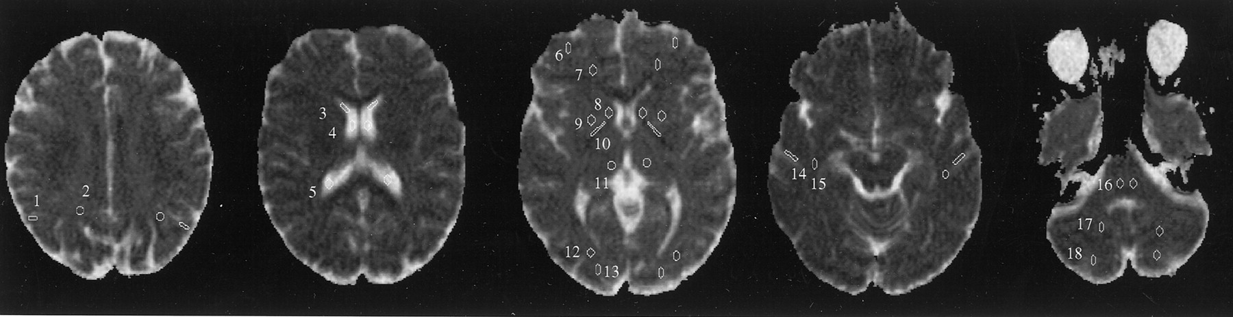
ROIs used in data analysis are superimposed on axial ADCav maps. 1 indicates the parietal gray matter; 2, parietal white matter; 3, frontal horn of lateral ventricle; 4, middle part of lateral ventricle; 5, posterior horn of lateral ventricle; 6, frontal gray matter; 7, frontal white matter; 8, caudate nucleus; 9, putamen; 10, internal capsule; 11, thalamus; 12, occipital white matter; 13, occipital gray matter; 14, temporal gray matter; 15, temporal white matter; 16, pons; 17, cerebellar white matter; and 18, cerebellar gray matter.
Statistical Analysis
The regional variation and the effect of sex on the ADCav values were studied by using the t test for dependent or independent samples, as appropriate. A one-way analysis of variance was applied to study the average values in the different regions of the brain. The groups representing different age cohorts and different sexes were compared with a multivariate analysis of variance. The homogeneity of the variances was studied with the Levene test. A two-tailed P value of less than .05 was considered to indicate a significant difference.
Results
In the whole brain, the mean ADCav values were (0.89 ± 0.04) × 10−3 mm2/s (range, 0.78–1.09 × 10⁻³ mm2/s) in the cortical gray matter, 0.70 ± 0.03 mm2/s (range, 0.62–0.79 × 10⁻³ mm2/s) in the white matter, (0.75 ± 0.03) × 10−3 mm2/s (range, 0.64–0.83 × 10⁻³ mm2/s) in the basal ganglia, and (0.73 ± 0.03) × 10−3 mm2/s (range, 0.67–0.82 × 10⁻³ mm2/s) in the thalamus. The regional ADCav values are presented in Tables 1 and 2.
ADCav values in the 36 ROIs
Mean ADCav values for gray matter from all the cerebral lobes compared with mean ADCav values for white matter
The mean cortical gray matter ADCav value was significantly higher than the mean white matter value in all subjects (P < .001), in men (P < .001), in women (P < .001), and in all age groups (P < .001). ADCav values in the basal ganglia and thalamus were higher than those in the white matter (P < .001), but they were lower than cortical gray matter values (P < .001) (Table 2). ADCav values in the white matter in the various regions of the brain did not differ significantly. No statistically significant differences were found between the various cortical regions. The range of ADCav values within each ROI was fairly large in all groups (Table 1). However, in every region and age group, the SDs of the ADCav values were small.
In both lateral ventricles, the ADCav values increased with age (P < .001). The homogeneity of the variances was normal in all the brain regions except the two thalami (P < .002 for the left and P < .001 for the right), where the ADCav values in age group 4 were significantly higher than those of the other age groups. No other statistically significant age-related changes in the ADCav values were noted. Differences in ADCav values between the hemispheres were found only in the cerebellar gray matter; the left hemisphere had a higher ADCav value than that of the right (P < .02). No differences were observed between the ADCav values in the men and those of the women.
Discussion
We report absolute ADCav values for the various regions of the human brain in a large representative adult population involving patients of both sexes with a wide age range. The study population was homogeneous; all subjects were white, and only two subjects were not Finnish. To our knowledge, no reports of normal ADC values in the brain in ethnically diverse populations exist, and whether our results can be directly extrapolated to people of different races requires further studies in such populations. Because the macro- and microstructural properties of the human brain are identical in people of various races, we believe that our results will prove to be valid for other populations as well.
The ADCav values in the cerebral cortex were consistently higher than those of the white matter; this finding supports the results of previous studies (12, 15, 21, 22). This difference is partly due to the structural and functional differences between the cortex and the white matter (21). The cortex has higher water content and substantially higher blood flow compared with that of the white matter (23); both contribute to the ADCav value. Furthermore, a partial volume effect of the CSF adjacent to the cortex may increase the ADCav values in the cortex (21, 22). In this study, the small variation among the ADCav values within a cortical ROI indicated that CSF contamination was avoided. As expected, the ADCav values in the CSF were more than twice as high as the gray matter values and almost three times as high as the ADCav values in the white matter, because water diffusion is much less restricted in the CSF than in the brain tissue. In previous reports, the ADC values in the gray matter were 0.8–1.1 × 10⁻³ mm2/s, and those in the white matter were 0.6–0.9 × 10⁻³ mm2/s (12, 22); these values are similar to our results. The ADCav values in the basal ganglia and thalamus were between the values in the gray matter and white matter, probably because of their microstructural properties (23).
Cerebellar gray matter ADCav values were higher in the left hemisphere than in the right hemisphere. In the cerebellum, avoiding some overlap between the gray matter ROIs and the white matter, and probably the CSF, is difficult, because the cerebellar cortex is thin and has extensive folia. This partial volume effect may explain the wide range of cerebellar ADCav values and the differences between the hemispheres.
Generally, the ADCav values in the various brain regions did not change with aging. However, an age-dependent increase in ADCav values was observed in the lateral ventricles, and the ADCav values in the thalami in age group 4 were significantly higher than in those of the other age groups. Whether the change in the thalami was due to structural or functional changes related to aging or mere statistical artifact remains to be shown. The change in the ADCav values in the lateral ventricles is possibly due to lower compliance in the aging brain and age-related enlargement of the ventricles, which allows more pulsatile movement of the CSF (24). This movement is likely to increase the turbulence of CSF flow (25), with a resultant increase in the measured diffusion (24). Gideon et al (15), in their study of 17 volunteers, found small age-related changes in the white matter but not in the gray matter. Other studies of ADC values in the healthy human brain have included only a few young subjects, or the total number of volunteers ranged between four and 15 (12, 16–18).
Absolute ADC values may be used to identify the ischemic tissue precisely (26), but their use requires a normal reference value for each brain region. Among human DW studies, only a few have involved the use of quantitative measurements (27–29). Quantitative methods, with absolute ADC values for healthy and pathologic structures, are important when focal and diffuse abnormalities are suspected, because minor changes may be difficult to detect by visual inspection. In experimental studies, comparisons of the ADC value in a given pixel before and after ischemia and the difference in ADC values in the infarcted and intact hemispheres have been used to detect early ischemia-induced ADC changes (1, 30–32). In humans, imaging before stroke is not feasible, and use of the contralateral side to provide a reference value may have limitations because of the possible existence of earlier lesions. A considerable amount of experimental work has been devoted to defining abnormal pixels and distinguishing normal pixels from ischemic pixels (26, 33–35). In studies of acute stroke, ADC values in the hyperacute (<6 hours after onset) ischemic lesion were less than 0.63 × 10⁻³ mm2/s (3, 36, 37). In this study, the mean ADCav values were (0.89 ± 0.04) × 10−3 mm2/s (range, 0.78–1.09 × 10⁻³ mm2/s) in the cortical gray matter, (0.70 ± 0.03) × 10−3 mm2/s (range, 0.62–0.79 × 10⁻³ mm2/s) in the white matter, and (0.75 ± 0.03) × 10−3 mm2/s (range, 0.64–0.83 × 10⁻³ mm2/s) in the basal ganglia; these results support the findings in hyperacute stroke studies. However, we identified some pixels with very low ADCav values; the lowest was 0.32 × 10⁻³ mm2/s, although most of the regional ADCav values were within a narrow range, and the calculated SDs were small. The rare pixels with surprisingly low ADCav values were randomly situated, and they may simply reflect blood vessels or technical errors. Because the ROIs were fairly large, these abnormal ADCav values did not affect the results.
ADC values alone are not site specific. As can be seen from the white matter ADCav values, no differences were found among the various brain regions. The same applies to the various cortical gray matter regions. Recent groups have indicated that knowledge of diffusion anisotropy can substantially improve the estimate for the anatomic location in question (38). In general, in the cerebral peduncles and internal capsule, the degree of diffusion anisotropy is high, whereas in the gray matter, diffusion is relatively isotropic (38–40). In the present study, the effects of anisotropy were overcome by using a rotationally invariant measure of diffusion (12).
Conclusion
In the present study, 80 healthy adult volunteers of both sexes with a wide age range were examined with spin-echo echo-planar DW imaging to establish reference normal average ADCav values in the various regions of the brain. With the exception of the lateral ventricles, the ADCav values were not affected by aging. The left and right hemispheres had similar ADCav values, and no sex difference was observed. In normal brain, the ADCav values in both gray matter and white matter were within a relatively narrow range. ADCav values beyond these boundaries may indicate disease, and thus, DW imaging may be useful for tissue characterization. Our data are representative, and our findings can be used for reference in future studies and in clinical settings.
Acknowledgments
We thank Dr Fuhai Li for his critical review of the manuscript.
Footnotes
Supported in part by the Maire Taponen Foundation, the University of Helsinki, the Sigrid Juselius Foundation, and the Foundation of Neurology, Helsinki, Finland.
References
- Received December 19, 2000.
- Accepted after revision September 26, 2001.
- Copyright © American Society of Neuroradiology
In this issue


Jump to section
Related Articles
Cited By...
- Freewater EstimatoR using iNtErpolated iniTialization (FERNET): Toward Accurate Estimation of Free Water in Peritumoral Region Using Single-Shell Diffusion MRI Data
- Diffusion-Weighted MR Imaging in a Prospective Cohort of Children with Cerebral Malaria Offers Insights into Pathophysiology and Prognosis
- Simulations of the effect of diffusion on asymmetric spin echo based quantitative BOLD: An investigation of the origin of deoxygenated blood volume overestimation
- Longitudinal diffusion changes following postoperative delirium in older people without dementia
- Early Prediction of Delayed Ischemia and Functional Outcome in Acute Subarachnoid Hemorrhage: Role of Diffusion Tensor Imaging
- A Simplified Model for Intravoxel Incoherent Motion Perfusion Imaging of the Brain
- The Effect of Age and Cerebral Ischemia on Diffusion-Weighted Proton MR Spectroscopy of the Human Brain
- Forensic Application of Postmortem Diffusion-Weighted and Diffusion Tensor MR Imaging of the Human Brain in Situ
- Mapping white matter diffusion and cerebrovascular reactivity in carotid occlusive disease
- Compromise of Brain Tissue Caused by Cortical Venous Reflux of Intracranial Dural Arteriovenous Fistulas: Assessment With Diffusion-Weighted Magnetic Resonance Imaging
- Impaired Cerebrovascular Reactivity With Steal Phenomenon Is Associated With Increased Diffusion in White Matter of Patients With Moyamoya Disease
- Neuroradiological characterization of normal adult ageing
- Usefulness of combined fractional anisotropy and apparent diffusion coefficient values for detection of involvement in multiple system atrophy
- Diffusion tensor magnetic resonance imaging at 3.0 tesla shows subtle cerebral grey matter abnormalities in patients with migraine
- Influence of aging on brain gray and white matter changes assessed by conventional, MT, and DT MRI
- Early diffusion weighted imaging and expression of heat shock protein 70 in newborn pigs with hypoxic ischaemic encephalopathy
- Magnetization transfer and diffusion tensor MRI show gray matter damage in neuromyelitis optica
- Brain diffusion changes in carotid occlusive disease treated with endarterectomy
- Longitudinal evaluation of leukoaraiosis with whole brain ADC histograms