Fig 3.
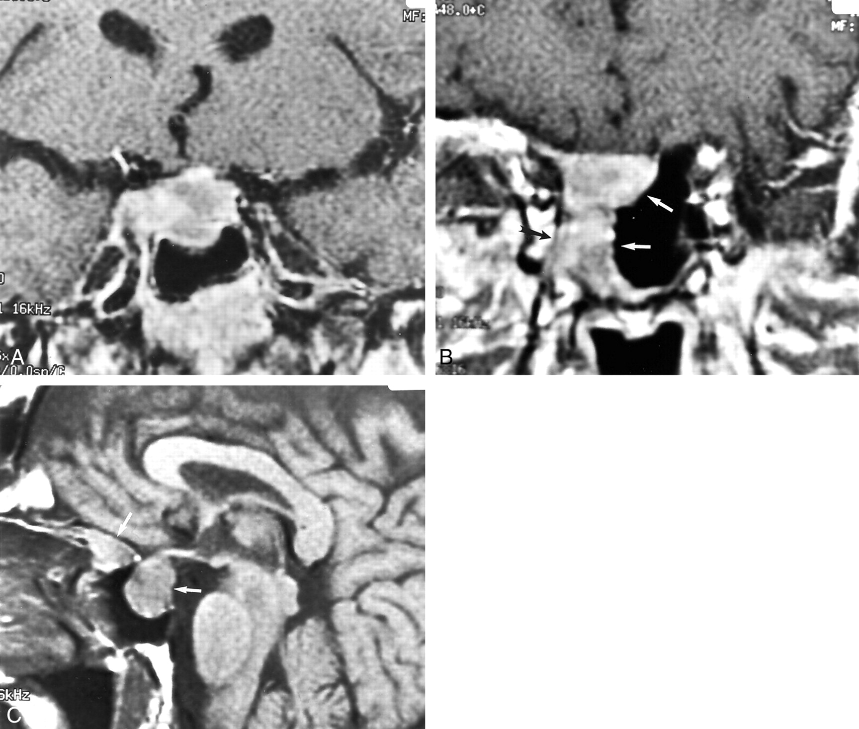
Fig 3.
Case 2. T1-weighted MR images of the sellar region.
A, Coronal image (600/15) depicts a heterogeneously enhancing sellar mass.
B, More anterior coronal section (600/15) depicts invasion of the right sphenoid sinus (white arrows) and cavernous sinus (black arrow).
C, Sagittal gadolinium-enhanced image (500/16) depicts two large portions of the sellar and parasellar mass (arrows).


