Fig 4.
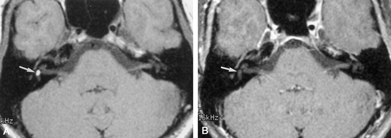
Fig 4.
Patient 4. (Images courtesy of Joel M. Schwartz, MD.)
A, Axial T1-weighted image (400/14/2) reveals a hyperintense mass in the right vestibule (arrow).
B, Right vestibular mass saturates on this T1-weighted axial fat-saturated image (500/20/4) (arrow), confirming a lipoma.


