Fig 1.

Fig 1.
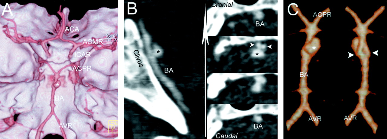
Scans from 16-row multisection CT and digital subtraction angiograms obtained before and after coiling.
A, 3D volume rendering of the multisection acquisition shows a panoramic view of the skull base with the position of the aneurysm (asterisk) at the middle segment of basilar artery.
B, Multiplanar reconstructions, obtained at the level of the clivus, show the position and configuration of the aneurysm and of the underlying basilar fenestration in the sagittal plane (left) and on sequential planes orthogonal to the clivus (right) through the basilar artery.
C, Segmented 3D volume rendering at the level of the basilar artery shows the aneurysm (craniocaudal view on left) and the fenestration (caudocranial view on right).


