Fig 4.

Fig 4.
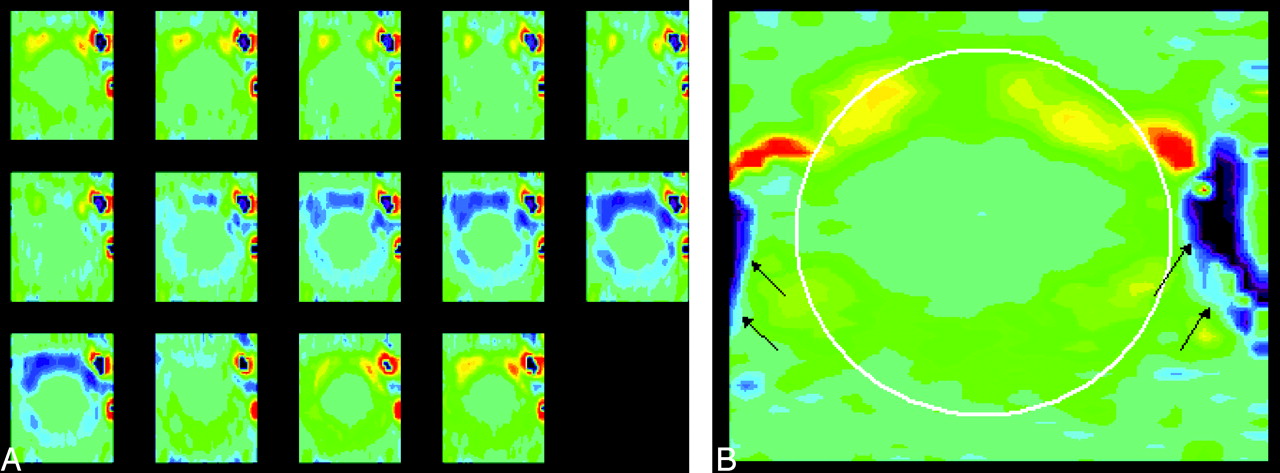
Representative phase contrast images of the foramen magnum in a patient (A) with the same vbgor color scale as in the normal subject. Flow velocities differ markedly in different regions in the subarachnoid space. Velocities anterior to the cord exceed those posterior to the cord. Velocities in the anterolateral subarachnoid space, exceed those elsewhere in the subarachnoid space, especially in diastole. A single frame (B) from late diastole is shown with a cursor placed to illustrate the region in which flow was measured (white oval) and the vertebral arteries (arrows). Note that flow in the subarachnoid space reverses while flow in the vertebral artery has continuous flow.


Related Articles
Cited By...
- Relationship between Cough-Associated Changes in CSF Flow and Disease Severity in Chiari I Malformation: An Exploratory Study Using Real-Time MRI
- Cough-Associated Changes in CSF Flow in Chiari I Malformation Evaluated by Real-Time MRI
- Current and Emerging MR Imaging Techniques for the Diagnosis and Management of CSF Flow Disorders: A Review of Phase-Contrast and Time-Spatial Labeling Inversion Pulse
- Physiology-Based MR Imaging Assessment of CSF Flow at the Foramen Magnum with a Valsalva Maneuver
- Patient-Specific 3D Simulation of Cyclic CSF Flow at the Craniocervical Region
- CSF Flow through the Upper Cervical Spinal Canal in Chiari I Malformation
- Peak CSF Velocities in Patients with Symptomatic and Asymptomatic Chiari I Malformation
- Accuracy and Reproducibility of Phase-Contrast MR Imaging Measurements for CSF Flow
- Characterization of CSF Hydrodynamics in the Presence and Absence of Tonsillar Ectopia by Means of Computational Flow Analysis
- Ethnic differences in syringomyelia in New Zealand
- Effect of Craniocervical Decompression on Peak CSF Velocities in Symptomatic Patients with Chiari I Malformation