Fig 3.

Fig 3.
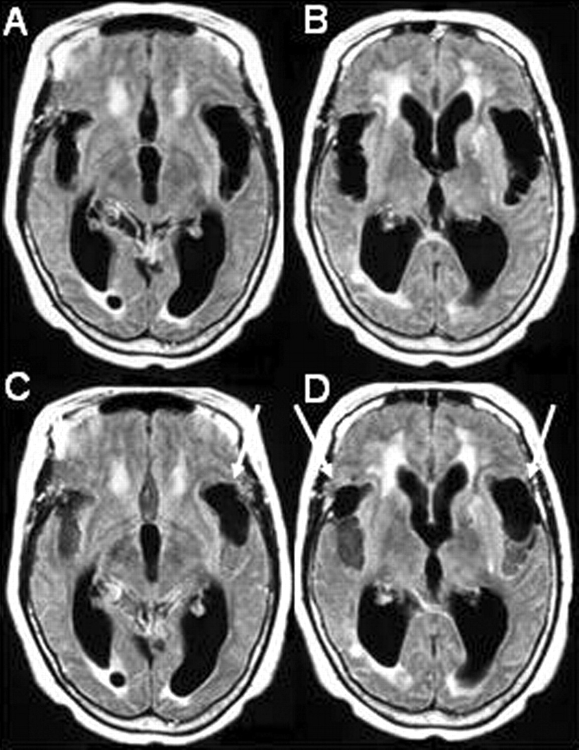
78-year-old female patient evaluated for suspected normal pressure hydrocephalus.
A and B, MRI FLAIR sequence (1.0 T, TR 11,000 ms, TE 140 ms, TI 2,600 ms) shows dilatation of the ventricles and Sylvian fissures.
C and D, FLAIR after 100% O2 for 5 minutes shows increased SI in the sulci and in the posterior aspects of both Sylvian fissures, allowing visualization of hypointense cysts (arrows) due to neurocysticerosis more anteriorly within the Sylvian fissures. Note that there is no increase in the SI of the CSF within the ventricles.


