Fig 2.

Fig 2.
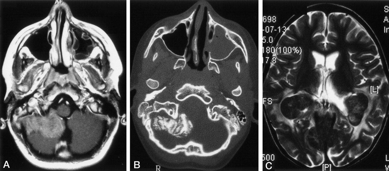
Images in two patients with LCH.
A and B, Lesions in a 13-year-old male patient at the diagnosis of LCH. Axial contrast-enhanced T1WI in A shows an extra-axial, enhancing, space-occupying lesion originating from the meninges. Axial bone-window CT scan in B shows calcification of the extra-axial lesion on the right side. Note the opacification of the left maxillary sinus.
C, Choriod plexus lesion in a 6-year-old girl with a 4.5-year history of LCH. Axial T2WIs show bilateral, hypointense masses in the choroid plexus and hyperintense changes in the parieto-occipital white matter.


Related Articles
Cited By...
- Neuroimaging in Pediatric Patients with Juvenile Xanthogranuloma of the CNS
- Waxing and Waning Neuroimaging Abnormalities in Langerhans Cell Histiocytosis
- The spectrum of immune-mediated and inflammatory lesions of the brainstem: Clues to diagnosis
- Cerebellar leukoencephalopathy: Most likely histiocytosis-related
- Neurodegenerative central nervous system disease as late sequelae of Langerhans cell histiocytosis. Report from the Japan LCH Study Group
- Improved outcome in multisystem Langerhans cell histiocytosis is associated with therapy intensification
- Differential diagnosis and evaluation in pediatric multiple sclerosis