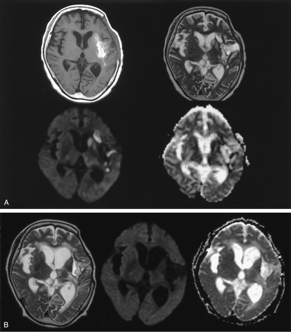
Fig 3.

Patient 8. A 66-year-old man with late subacute intracerebral hematoma on MR images obtained 30 days after symptom onset.
A, T1-weighted image (top left) shows hyperintensity of the hematoma mainly in the left external capsule area. Hematoma also extends into a part of the posterior angle of the putamen and insular cortex. T2-weighted image (top right) shows the hematoma as a hyperintense area circumscribed by a hypointense rim; this suggests subacute hematoma. Subtle hyperintensity is noted in the ipsilateral caudate head and putamen. DW image (bottom left) shows obvious hyperintensity in the caudate and putamen and mild hyperintensity in the thalamus. ADC map (bottom right) shows decreased ADC in the caudate, part of the putamen and medial-posterior thalamus; ADC ratios relative to the contralateral gray matter were 0.59, 0.61, and 0.87, respectively.
B, T2-weighted image (left) obtained 12 months later shows atrophy of the caudate and putamen, while the hematoma resolves to a slitlike cavity. Follow-up diffusion image (middle) reveals no definable signal intensity change in the caudate, putamen, or thalamus ipsilateral to the hematoma. Follow-up ADC map (right) shows normalization of ADC values in the corresponding deep gray matter.


