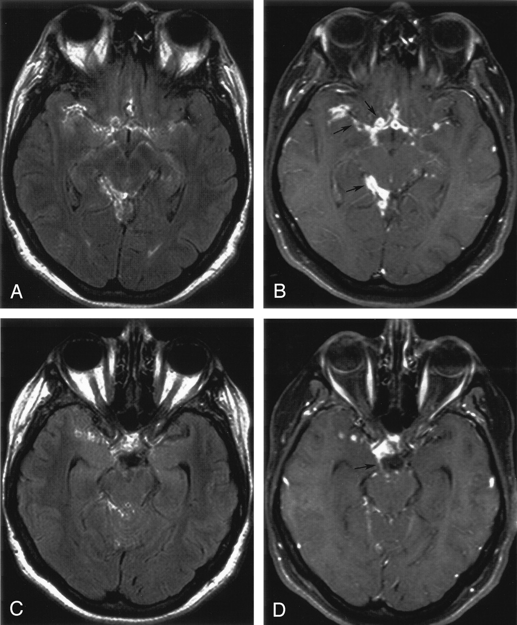
Fig 2.

Tuberculous meningitis.
A, Contrast-enhanced FLAIR image shows mildly enhancing subarachnoid space lesions in the basilar cisterns, with extension into the sylvian fissures bilaterally, right ambient cistern, and quadrigeminal cistern.
B, Contrast-enhanced T1-weighted image with FS shows the enhancing lesions in the subarachnoid space more definitively (arrows). The contrast difference between the enhancing meninges and the adjacent brain is visually greater in the T1-weighted image.
C, Contrast-enhanced FLAIR image of the same patient shows enhancement in the region of the right third cranial nerve, but the nerve is not clearly seen.
D, The enhancement of the right third cranial nerve (arrow) is more distinct on the contrast-enhanced T1-weighted image with FS.


Related Articles
Cited By...
- Diagnostic Accuracy of MRI for Detection of Meningitis in Infants
- Potentially Reversible and Recognizable Acute Encephalopathic Syndromes: Disease Categorization and MRI Appearances
- Comparison of the Added Value of Contrast-Enhanced 3D Fluid-Attenuated Inversion Recovery and Magnetization-Prepared Rapid Acquisition of Gradient Echo Sequences in Relation to Conventional Postcontrast T1-Weighted Images for the Evaluation of Leptomeningeal Diseases at 3T