
For several years now, radiologists have known the utility of diffusion-weighted (DW) imaging in differentiating brain abscesses from cystic/necrotic brain tumors.
In “Diffusion-Weighted Imaging in the Assessment of Brain Abscesses Therapy,” Cartes-Zumelzu et al (1) discuss the extension of this technique to using DW imaging to assess abscess response to therapy. In the article, the authors state, “with contrast-enhanced T1-weighted imaging the differentiation of purulent fluid from serous fluid inside the abscess cavity is not possible.” They further conclude, “findings in successfully and unsuccessfully treated brain abscesses were similar on conventional MR images and did not allow for their differentiation.” In my experience this is not always the case. Clearly, DW imaging is a very useful technique to assess the therapeutic response of the abscess cavity, however this may also be done by careful inspection of routine contrast-enhanced T1-weighted images. I have included an example to illustrate this point. The images presented are from a child of 3 weeks of age with Gram-negative rod meningitis.
On the initial images (Figs 1–3) multiple abscesses were noted in both frontal and parietal lobes bilaterally, which were homogeneously low (but not CSF) signal intensity with peripheral enhancement on postcontrast T1-weighted images. They appeared hyperintense on DW imaging and demonstrated corresponding hypointensity on the apparent diffusion coefficient (ADC) map.
Following 11 days of antibiotic therapy (ampicillin and gentamicin) the patient was reimaged (Figs 4–6). Again ring-enhancing lesions were noted on the postcontrast T1-weighted images. But, unlike the initial images, the collections were heterogeneous with two discrete components. The dependent portion was again hypointense (but not CSF in signal intensity) and again demonstrated restricted diffusion. The nondependent portion, however, became isointense to CSF and was no longer hyperintense on DW imaging and was hypointense on the ADC map. Thus, the postcontrast T1-weighted images demonstrated essentially the same information (albeit a bit more subtle) as the DW image. This in no way diminishes the use of diffusion sequences in diagnosing and following brain abscesses, but merely points out the usefulness of conventional T1-weighted images.

Axial contrast-enhanced T1-weighted image.
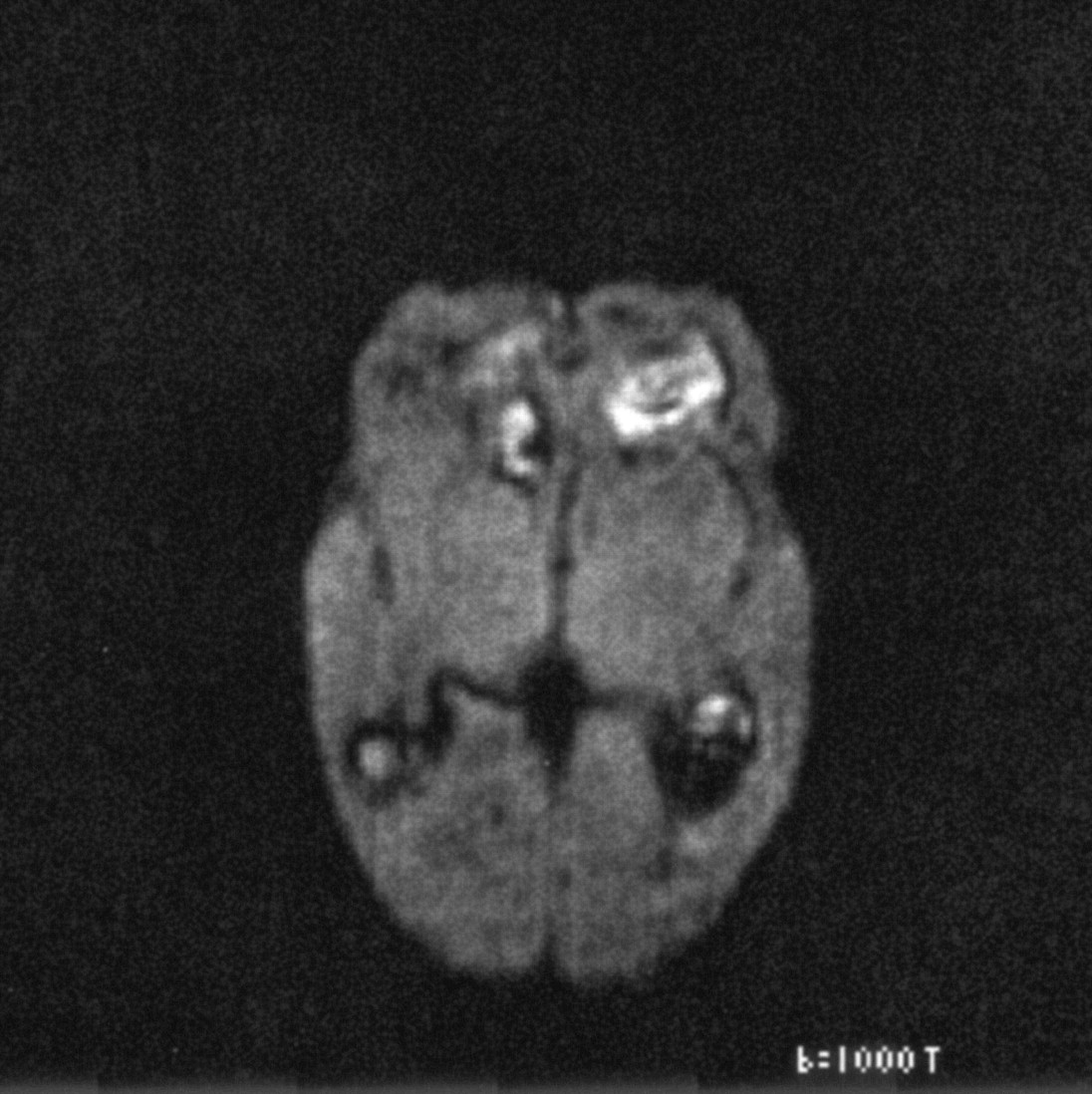
Axial DW image.

Axial ADC map.

Axial contrast-enhanced T1-weighted image.

Axial DW image.
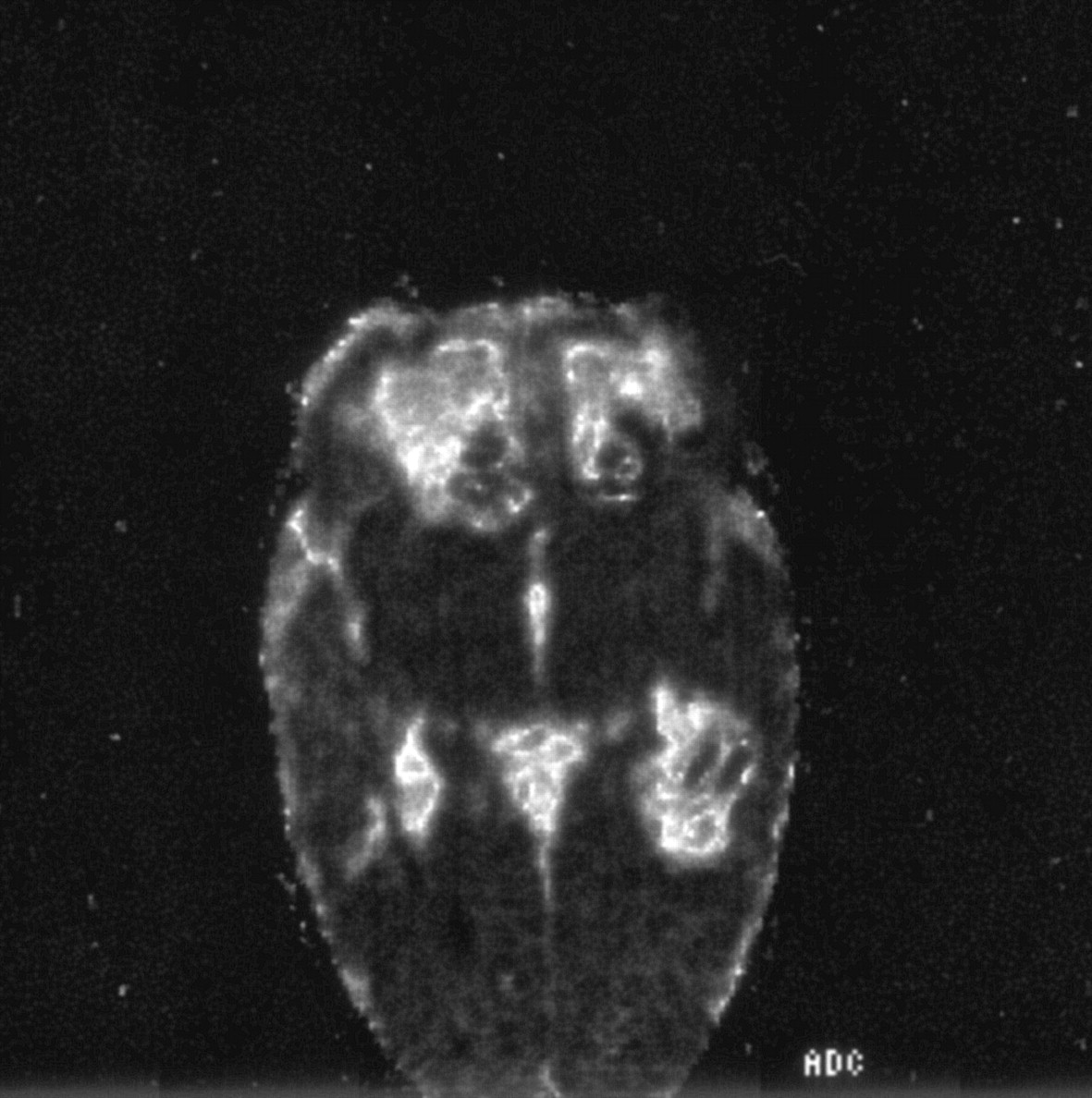
Axial ADC image.
Reference
- Copyright © American Society of Neuroradiology
In this issue


Jump to section
Related Articles
Cited By...
- No citing articles found.