Fig 2.
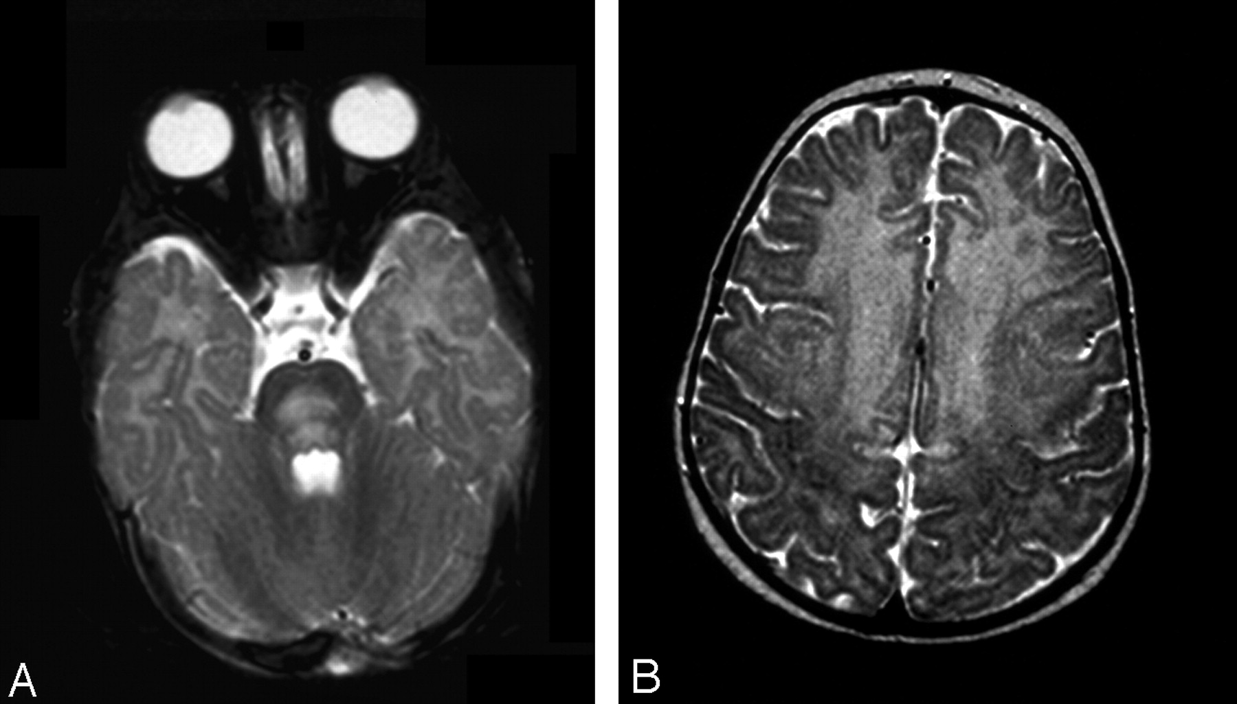
Fig 2.
Patient 2. Axial T2-weighted images.
A, At presentation, large area of hyperintensity is present in the pons.
B, Involvement of the frontal region, up to the motor cortex, is extensive.


Related Articles
Cited By...
- Signal Change in the Mammillary Bodies after Perinatal Asphyxia
- Cranial Ultrasonography in Infantile Encephalitic Beriberi: A Useful First-Line Imaging Tool for Screening and Diagnosis in Suspected Cases
- Infantile Wernicke's encephalopathy
- TEACHING NEUROIMAGES: THE FULL-BLOWN NEUROIMAGING OF WERNICKE ENCEPHALOPATHY
- Epilepsy in children with infantile thiamine deficiency