Fig 6.
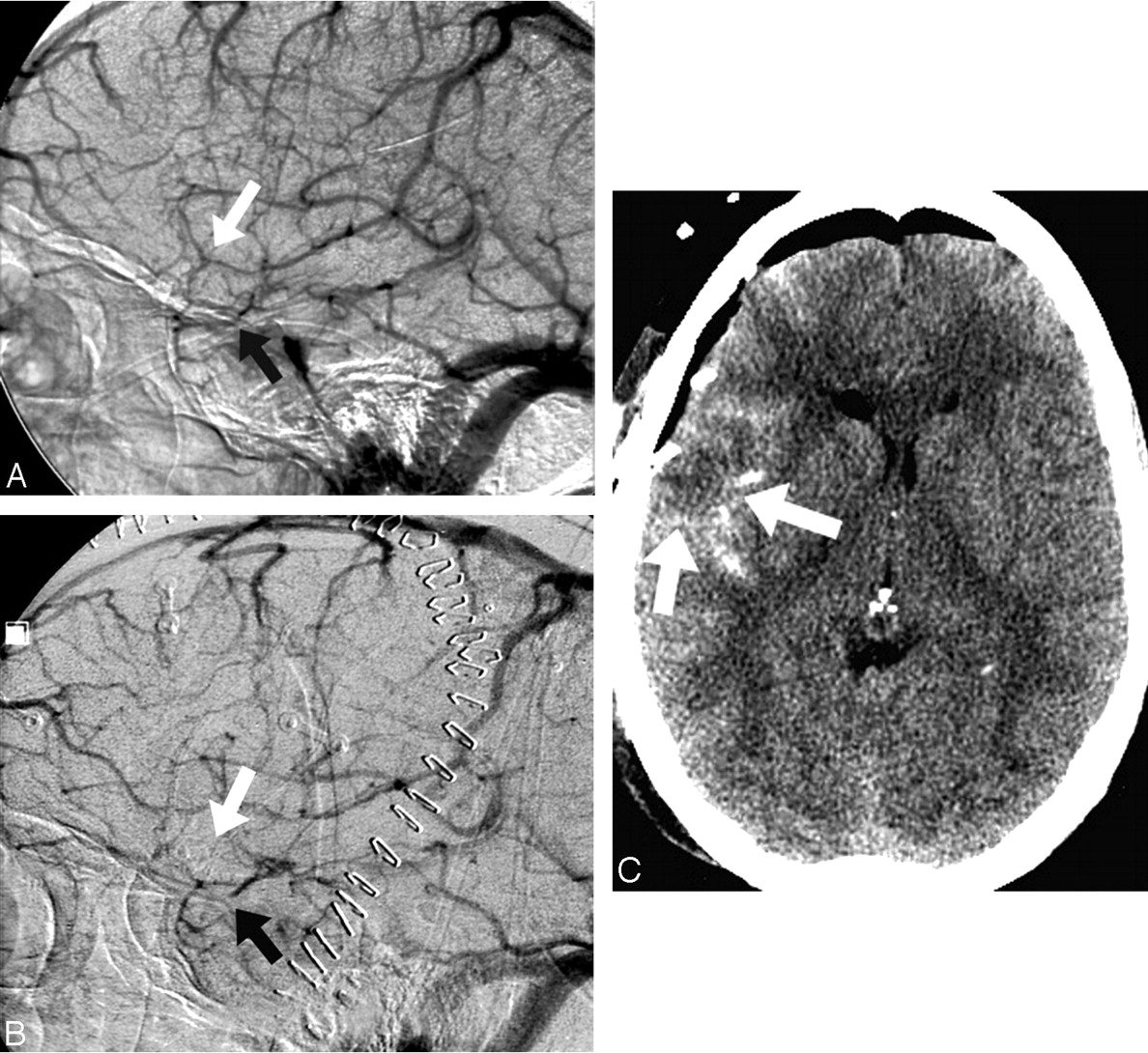
Fig 6.
Occluded and very small SMCV and small temporal branch are associated with a small region of brain edema. A, Very small SMCV (white arrow); temporal branch (black arrow). B, Nonfilling of tiny SMCV (white arrow) and distal occlusion of temporal branch (black arrow). C, Small temporal lobe region of edema appears to correlate well with small vessel occlusions (arrows).


