Fig 1.
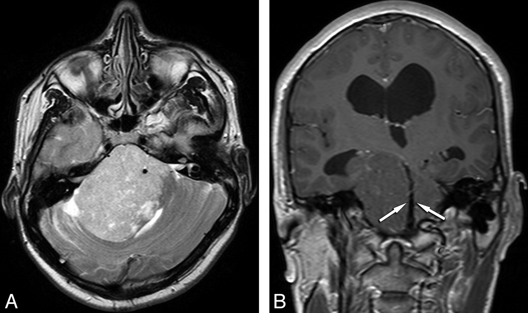
Fig 1.
A, Axial T2-weighted image shows a large, solid hyperintense mass in the right CPA and prepontine cistern, displacing the fourth ventricle to left. The signal intensity void of basilar artery is maintained but is entirely circumscribed by tumor. B, Coronal contrast-enhanced T1-weighted image shows the patent basilar artery (arrows) and obstructive hydrocephalus. On the left, the tumor traverses the tentorial incisura and displaces the third ventricle to the left. There is minimal punctate enhancement.


Related Articles
Cited By...
- No citing articles found.