Fig 2.

Fig 2.
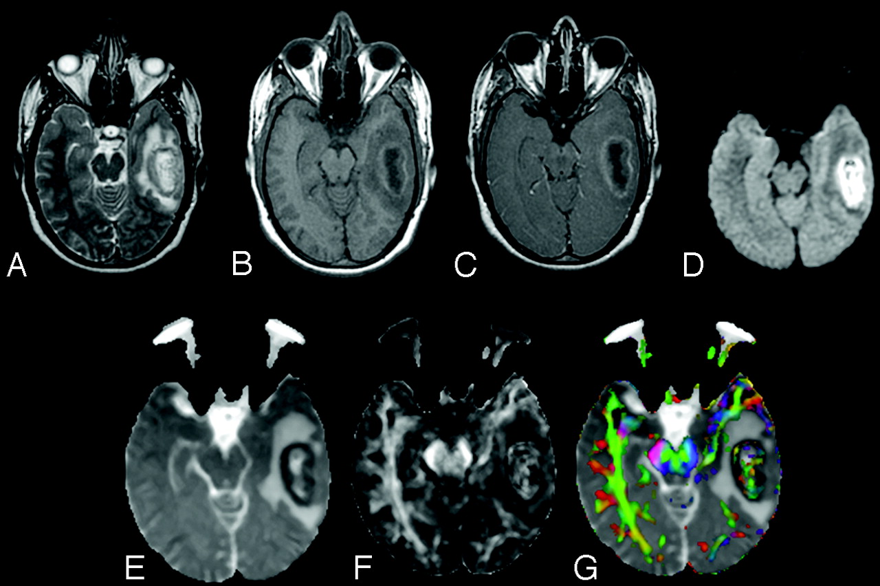
A 25-year-old male patient presenting with pyogenic brain abscess with a heterogeneously hyperintense lesion in the left temporal lobe. A, Axial T2-weighted image shows a hyperintense lesion with a hypointense wall. B, The lesion appears hypointense on the axial T1-weighted image with an isointense wall. C, On the postcontrast T1-weighted image, the lesion shows ring enhancement. D, The lesion appears heterogeneously hyperintense on the DWI in the cavity that appears heterogeneously hypointense on the MD map (E). The FA (F) and red-green-blue color-modulated FA map fused with MD map (G) show that high FA in the abscess cavity is similar to what is observed in the contralateral inferior longitudinal fasciculus and midbrain.


Related Articles
Cited By...
- Differential Gene Expression in Glioblastoma Defined by ADC Histogram Analysis: Relationship to Extracellular Matrix Molecules and Survival
- Differentiation of Tumefactive Demyelinating Lesions from High-Grade Gliomas with the Use of Diffusion Tensor Imaging
- Apparent Diffusion Coefficient with Higher b-Value Correlates Better with Viable Cell Count Quantified from the Cavity of Brain Abscess
- Differentiation of Brain Abscesses from Necrotic Glioblastomas and Cystic Metastatic Brain Tumors with Diffusion Tensor Imaging
- Correlation of Quantitative Diffusion Tensor Tractography with Clinical Grades of Subacute Sclerosing Panencephalitis
- In Vivo Proton MR Spectroscopy Evaluation of Pyogenic Brain Abscesses: A Report of 194 Cases