Fig 3.
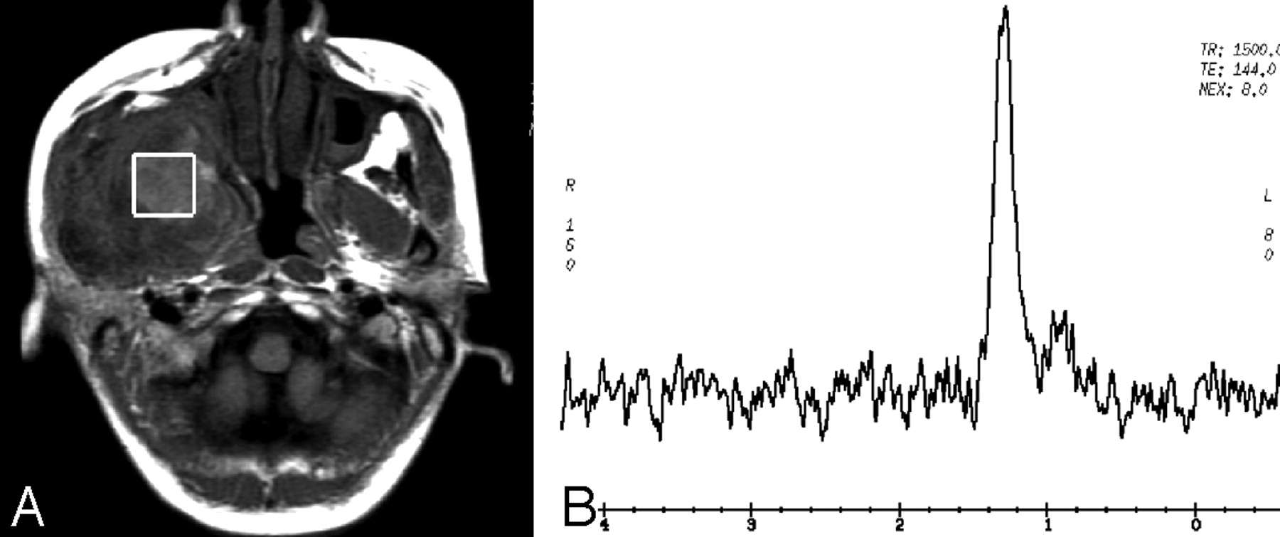
Fig 3.
Case 1 with a chronic inflammatory lesion in the right masticator space. Axial T1-weighted image (A) demonstrates that the lesion with isointense signal intensity is located in the right medial masticator space. The box of VOI is positioned in the lesion of the right medial masticator space, and there are no Cho signals present on the spectrum (B). The lipid signals are shown at 0.9–1.5 ppm.


