Fig 3.
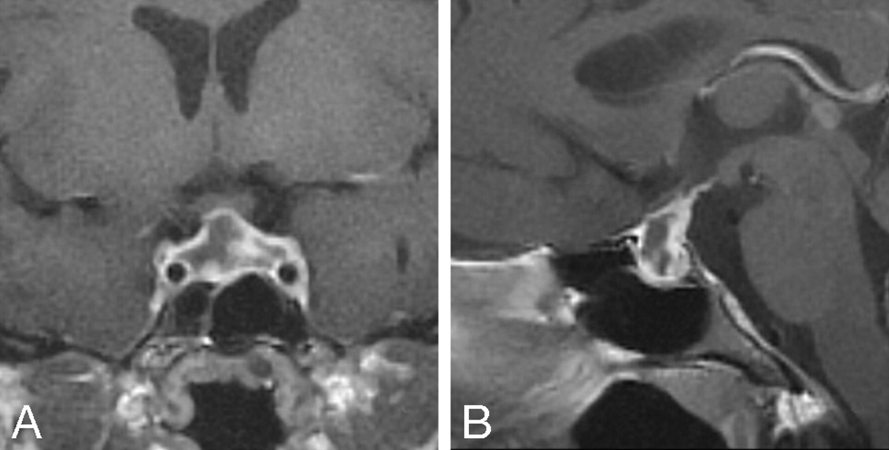
Fig 3.
Case 2. Coronal (A) and sagittal (B) gadolinium-enhanced T1-weighted MR images on postpartum day 6 show a large low signal intensity sellar lesion with enhancement of the rim. There is extension into the suprasellar cistern, and the lesion abuts but does not compress the optic chiasm.


