Fig 1.

Fig 1.
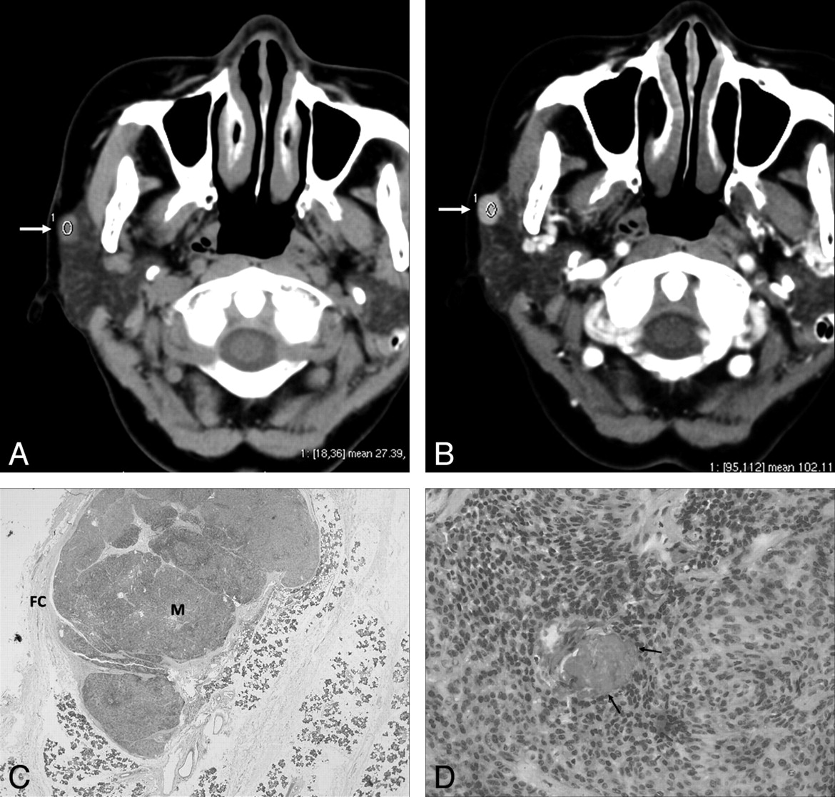
A 62-year-old woman with ME. A, Precontrast axial CT scan shows a round, well-defined mass (27.39 HU) in the superficial lobe of the right parotid gland abutting on the capsule of gland (arrow). B, Contrast-enhanced axial CT scan shows the mass (102.11 HU) with homogeneous strong enhancement (arrow). C, Microscopic examination (hematoxylin-eosin stain [HE]; ×2) shows that the ME (M) is separated from the parotid gland and the surrounding soft tissues by a fibrous capsule (FC). The stroma is scanty. D, Microscopic examination (HE; ×100) shows that the tumor is composed of the plasmacytoid and spindle cells. Relatively large vessels are seen in the tumor (arrows).


Related Articles
Cited By...
- No citing articles found.