Advertisement

Main menu

  • Home
  • Content
    • Current Issue
    • Publication Preview--Ahead of Print
    • Past Issue Archive
    • Case of the Week Archive
    • Classic Case Archive
    • Case of the Month Archive
  • For Authors
  • About Us
    • About AJNR
    • Editors
    • American Society of Neuroradiology
  • Submit a Manuscript
  • Podcasts
    • Subscribe on iTunes
    • Subscribe on Stitcher
  • More
    • Subscribers
    • Permissions
    • Advertisers
    • Alerts
    • Feedback
  • Other Publications
    • ajnr

User menu

  • Subscribe
  • Alerts
  • Log in

Search

  • Advanced search
American Journal of Neuroradiology

American Journal of Neuroradiology

  • Subscribe
  • Alerts
  • Log in

Advanced Search

  • Home
  • Content
    • Current Issue
    • Publication Preview--Ahead of Print
    • Past Issue Archive
    • Case of the Week Archive
    • Classic Case Archive
    • Case of the Month Archive
  • For Authors
  • About Us
    • About AJNR
    • Editors
    • American Society of Neuroradiology
  • Submit a Manuscript
  • Podcasts
    • Subscribe on iTunes
    • Subscribe on Stitcher
  • More
    • Subscribers
    • Permissions
    • Advertisers
    • Alerts
    • Feedback
  • Follow AJNR on Twitter
  • Visit AJNR on Facebook
  • Follow AJNR on Instagram
  • Join AJNR on LinkedIn
  • RSS Feeds
Research ArticleBrain

Acute Stroke Evaluated by Time-to-Peak Mapping during Initial and Early Follow-up Perfusion CT Studies

Jürgen R. Reichenbach, Joachim Röther, Lars Jonetz-Mentzel, Michael Herzau, Anke Fiala, Cornelius Weiller and Werner A. Kaiser
American Journal of Neuroradiology November 1999, 20 (10) 1842-1850;
Jürgen R. Reichenbach
  • Find this author on Google Scholar
  • Find this author on PubMed
  • Search for this author on this site
Joachim Röther
  • Find this author on Google Scholar
  • Find this author on PubMed
  • Search for this author on this site
Lars Jonetz-Mentzel
  • Find this author on Google Scholar
  • Find this author on PubMed
  • Search for this author on this site
Michael Herzau
  • Find this author on Google Scholar
  • Find this author on PubMed
  • Search for this author on this site
Anke Fiala
  • Find this author on Google Scholar
  • Find this author on PubMed
  • Search for this author on this site
Cornelius Weiller
  • Find this author on Google Scholar
  • Find this author on PubMed
  • Search for this author on this site
Werner A. Kaiser
  • Find this author on Google Scholar
  • Find this author on PubMed
  • Search for this author on this site
  • Article
  • Figures & Data
  • Info & Metrics
  • References
  • PDF
Loading

Article Figures & Data

Figures

  • Tables
  • fig 1.
    • Download figure
    • Open in new tab
    • Download powerpoint
    fig 1.

    Patient 6.

    A, Initial native CT scan shows standardized circular ROIs, which were manually placed in both hemispheres in the vascular territories of the ACA, MCA, PCA, the basal ganglia region, and the thalamus.

    B, Enhancement-versus-time curves of ROIs 1 and 2 located symmetrically in the anterior vascular territory of the MCA in the ischemic and healthy areas, respectively. Note the strong enhancement peak indicating the passage of the compact contrast bolus in the normal area (ROI 2). Conversely, the time course of the contralateral ROI (ROI 1) shows a markedly delayed and slow enhancement, indicating hypoperfusion.
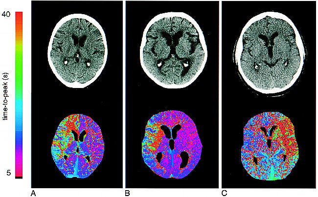
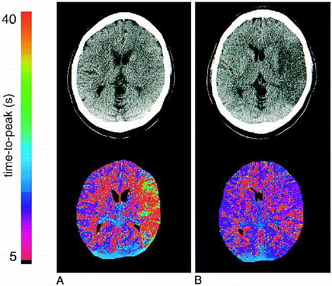
  • fig 2.
    • Download figure
    • Open in new tab
    • Download powerpoint
    fig 2.

    Initial native CT scans and corresponding time-to-peak parameter maps. The maps are color-coded with values ranging from 5 to 40 seconds.

    A, Patient 6: 62-year-old woman who presented 40 minutes after onset of left hemiparesis. The CT scan is normal. Note the clear demarcation of the diseased area in the right hemisphere, corresponding to the right ACA and MCA territories, including the basal ganglia on the parameter map.

    B, Patient 17: 77-year-old woman who presented 40 minutes after onset of somnolence and left hemiparesis. The native CT scan is normal. The parameter map shows an area of delayed perfusion in the territory of the right MCA.

    C, Patient 1: 60-year-old man who had a normal CT scan 2.8 hours after onset. The time-to-peak map depicts a large perfusion deficit in the left MCA territory, including the basal ganglia.

  • fig 3.
    • Download figure
    • Open in new tab
    • Download powerpoint
    fig 3.

    Patient 10: initial and follow-up examinations.

    A, 2 hours 50 minutes after onset.

    B, 30 hours after onset, during therapy with heparin. The patient suffered from a large infarct in the left hemisphere with global aphasia and right hemiparesis. The first native CT scan (A) reveals only discrete cortical hypodensity and effacement of cortical sulci in the ischemic hemisphere. In contrast, the time-to-peak map clearly shows the extent of the affected brain area, which corresponds strongly to the hypodensity seen on the CT scan in the follow-up study (B). The time-to-peak map shows tissue reperfusion 30 hours later. A large MCA infarction developed despite recanalization.

Tables

  • Figures
  • TABLE 1:
    • View popup
    • Download powerpoint
    TABLE 1:

    Clinical data of patients

  • TABLE 2:
    • View popup
    • Download powerpoint
    TABLE 2:

    Clinical data and hemispheric lesion areas (HLA) during initial and follow-up scans

PreviousNext
Back to top

In this issue

American Journal of Neuroradiology
Vol. 20, Issue 10
1 Nov 1999
  • Table of Contents
  • Index by author
Advertisement
Print
Download PDF
Email Article

Thank you for your interest in spreading the word on American Journal of Neuroradiology.

NOTE: We only request your email address so that the person you are recommending the page to knows that you wanted them to see it, and that it is not junk mail. We do not capture any email address.

Enter multiple addresses on separate lines or separate them with commas.
Acute Stroke Evaluated by Time-to-Peak Mapping during Initial and Early Follow-up Perfusion CT Studies
(Your Name) has sent you a message from American Journal of Neuroradiology
(Your Name) thought you would like to see the American Journal of Neuroradiology web site.
CAPTCHA
This question is for testing whether or not you are a human visitor and to prevent automated spam submissions.
Citation Tools
Acute Stroke Evaluated by Time-to-Peak Mapping during Initial and Early Follow-up Perfusion CT Studies
Jürgen R. Reichenbach, Joachim Röther, Lars Jonetz-Mentzel, Michael Herzau, Anke Fiala, Cornelius Weiller, Werner A. Kaiser
American Journal of Neuroradiology Nov 1999, 20 (10) 1842-1850;

Citation Manager Formats

  • BibTeX
  • Bookends
  • EasyBib
  • EndNote (tagged)
  • EndNote 8 (xml)
  • Medlars
  • Mendeley
  • Papers
  • RefWorks Tagged
  • Ref Manager
  • RIS
  • Zotero
Share
Acute Stroke Evaluated by Time-to-Peak Mapping during Initial and Early Follow-up Perfusion CT Studies
Jürgen R. Reichenbach, Joachim Röther, Lars Jonetz-Mentzel, Michael Herzau, Anke Fiala, Cornelius Weiller, Werner A. Kaiser
American Journal of Neuroradiology Nov 1999, 20 (10) 1842-1850;
del.icio.us logo Digg logo Reddit logo Twitter logo CiteULike logo Facebook logo Google logo Mendeley logo
  • Tweet Widget
  • Facebook Like
  • Google Plus One

Jump to section

  • Article
    • Abstract
    • Methods
    • Results
    • Discussion
    • Conclusion
    • Footnotes
    • References
  • Figures & Data
  • Info & Metrics
  • References
  • PDF

Related Articles

  • No related articles found.
  • PubMed
  • Google Scholar

Cited By...

  • Perfusion Deficits and Mismatch in Patients with Acute Lacunar Infarcts Studied with Whole-Brain CT Perfusion
  • Evaluation of CT Perfusion in the Setting of Cerebral Ischemia: Patterns and Pitfalls
  • Perfusion CT in Acute Ischemic Stroke: A Qualitative and Quantitative Comparison of Deconvolution and Maximum Slope Approach
  • Parametric Color Coding of Digital Subtraction Angiography
  • Fetal Origin of the Posterior Cerebral Artery Produces Left-Right Asymmetry on Perfusion Imaging
  • Reproducibility of Quantitative CT Brain Perfusion Measurements in Patients with Symptomatic Unilateral Carotid Artery Stenosis
  • Perfusion-CT Assessment of Infarct Core and Penumbra: Receiver Operating Characteristic Curve Analysis in 130 Patients Suspected of Acute Hemispheric Stroke
  • Visual evaluation of perfusion computed tomography in acute stroke accurately estimates infarct volume and tissue viability
  • Accuracy of Dynamic Perfusion CT with Deconvolution in Detecting Acute Hemispheric Stroke
  • Prediction of Neurologic Deterioration in Patients with Lacunar Infarction in the Territory of the Lenticulostriate Artery Using Perfusion CT
  • Bedside assessment of cerebral perfusion reductions in patients with acute ischaemic stroke by near-infrared spectroscopy and indocyanine green
  • Correlation of Early Dynamic CT Perfusion Imaging with Whole-Brain MR Diffusion and Perfusion Imaging in Acute Hemispheric Stroke
  • Trial Design and Reporting Standards for Intra-Arterial Cerebral Thrombolysis for Acute Ischemic Stroke
  • Safety and Feasibility of a CT Protocol for Acute Stroke: Combined CT, CT Angiography, and CT Perfusion Imaging in 53 Consecutive Patients
  • Guidelines and Recommendations for Perfusion Imaging in Cerebral Ischemia: A Scientific Statement for Healthcare Professionals by the Writing Group on Perfusion Imaging, From the Council on Cardiovascular Radiology of the American Heart Association
  • Computed Tomographic Findings in Patients Undergoing Intra-arterial Thrombolysis for Acute Ischemic Stroke due to Middle Cerebral Artery Occlusion: Results From the PROACT II Trial * Editorial Comment: Results From the PROACT II Trial
  • Perfusion CT Scans
  • Multisection Dynamic CT Perfusion for Acute Cerebral Ischemia: The "Toggling-table" Technique
  • Correlation of Cerebrovascular Reserve as Measured by Acetazolamide-challenged SPECT with Angiographic Flow Patterns and Intra- or Extracranial Arterial Stenosis
  • Quantitative Assessment of the Ischemic Brain by Means of Perfusion-Related Parameters Derived From Perfusion CT
  • Intraarterial Thrombolysis: Ready for Prime Time?
  • Perfusion Mapping Using Computed Tomography Allows Accurate Prediction of Cerebral Infarction in Experimental Brain Ischemia Editorial Comment
  • Crossref
  • Google Scholar

This article has not yet been cited by articles in journals that are participating in Crossref Cited-by Linking.

More in this TOC Section

  • Progression of Microstructural Damage in Spinocerebellar Ataxia Type 2: A Longitudinal DTI Study
  • SWI or T2*: Which MRI Sequence to Use in the Detection of Cerebral Microbleeds? The Karolinska Imaging Dementia Study
  • Statin Therapy Does Not Affect the Radiographic and Clinical Profile of Patients with TIA and Minor Stroke
Show more BRAIN

Similar Articles

Advertisement

News and Updates

  • Lucien Levy Best Research Article Award
  • Thanks to our 2020 Distinguished Reviewers
  • Press Releases

Resources

  • Evidence-Based Medicine Level Guide
  • How to Participate in a Tweet Chat
  • AJNR Podcast Archive
  • Ideas for Publicizing Your Research
  • Librarian Resources
  • Terms and Conditions

Opportunities

  • Share Your Art in Perspectives
  • Get Peer Review Credit from Publons
  • Moderate a Tweet Chat

American Society of Neuroradiology

  • Neurographics
  • ASNR Annual Meeting
  • Fellowship Portal
  • Position Statements

© 2021 by the American Society of Neuroradiology | Print ISSN: 0195-6108 Online ISSN: 1936-959X

Powered by HighWire