
Article Figures & Data
Figures
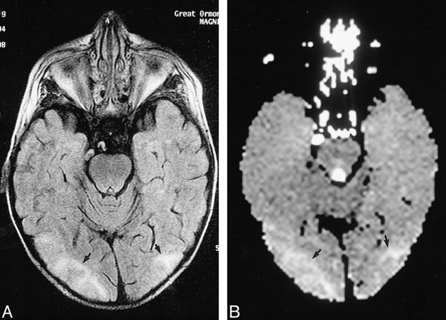
- fig 1.
Axial MR images of a 4-year-old patient (case 1) obtained on day 30 after bone marrow transplantation. The patient presented with lethargy and focal seizures secondary to CSA therapy. The T2 signal abnormalities corresponded to areas of increased diffusion, suggesting that these areas of cerebral edema were caused by extravasation of fluid and not cerebral ischemia.
A, A fluid-attenuated inversion-recovery image shows confluent cortical and juxtacortical signal alteration in both occipital lobes (arrows).
B, The ADC map at the same level reveals increased diffusion in the regions of prolonged T2 signal (arrows). (Normal brain = 1.00 × 10⁻³ mm2/second, abnormal areas = 1.42.)
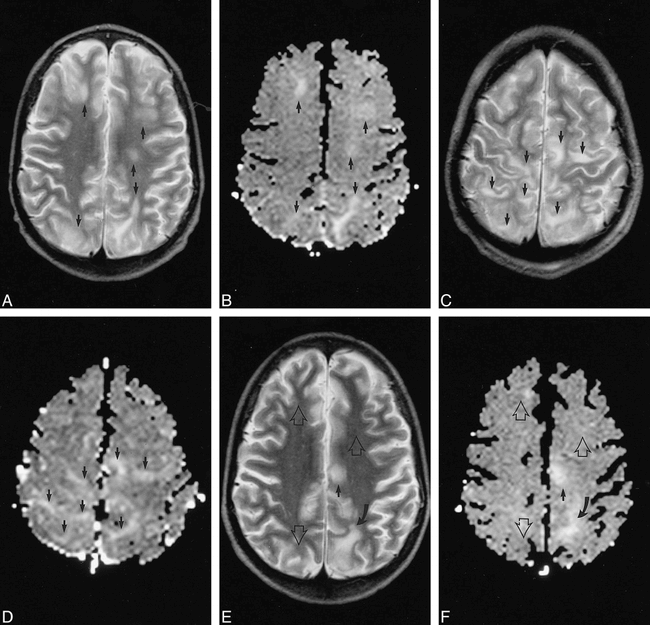
- fig 2.
Axial MR images of a 9-year-old patient (case 2) with sickle cell disease and a viral-induced nephrotic syndrome. The patient developed a generalized seizure 4 days after commencing CSA therapy and experienced prolonged impairment of higher neurologic function. Regions of increased diffusion matched all the areas of T2 signal change. Many of the areas resolved without any residual T2 or diffusion abnormality. These findings suggest that the neurotoxic effects of CSA were associated with a partially reversible extravasation of fluid into the brain.
A, A T2-weighted image (day 2 after seizure) shows cortical and juxtacortical signal alteration within both frontal and parietal lobes (arrows).
B, The ADC map (day 2) reveals increased diffusion in all areas of T2 abnormality (arrows). (Normal brain = 1.02, abnormal areas = 1.33.)
C, A T2-weighted image (day 6) shows new, more superior abnormalities (arrows).
D, The ADC map (day 6) again reveals that all of the T2-weighted abnormalities correspond to areas of increased diffusion (arrows). (Normal brain = 0.93, abnormal areas = 1.48.)
E, A T2-weighted image (day 49), obtained at the same level as the images presented in A and B, shows complete resolution of most of the lesions that were present on day 2 (open arrows). A new lesion is present in the left posterior frontal lobe (solid arrow). A preexisting abnormality in the left parietal lobe (curved arrow) is essentially unchanged.
F, The ADC map (day 49) reveals increased diffusion in the new area of T2 prolongation (solid arrow). No diffusion abnormality, however, is present where the lesions have resolved (open arrows). (Normal brain = 0.93, open arrows = 0.92, solid arrow = 1.34, curved arrow = 1.36.)


