Advertisement

Main menu

  • Home
  • Content
    • Current Issue
    • Publication Preview--Ahead of Print
    • Past Issue Archive
    • Case of the Week Archive
    • Classic Case Archive
    • Case of the Month Archive
  • For Authors
  • About Us
    • About AJNR
    • Editors
    • American Society of Neuroradiology
  • Submit a Manuscript
  • Podcasts
    • Subscribe on iTunes
    • Subscribe on Stitcher
  • More
    • Subscribers
    • Permissions
    • Advertisers
    • Alerts
    • Feedback
  • Other Publications
    • ajnr

User menu

  • Subscribe
  • Alerts
  • Log in

Search

  • Advanced search
American Journal of Neuroradiology

American Journal of Neuroradiology

  • Subscribe
  • Alerts
  • Log in

Advanced Search

  • Home
  • Content
    • Current Issue
    • Publication Preview--Ahead of Print
    • Past Issue Archive
    • Case of the Week Archive
    • Classic Case Archive
    • Case of the Month Archive
  • For Authors
  • About Us
    • About AJNR
    • Editors
    • American Society of Neuroradiology
  • Submit a Manuscript
  • Podcasts
    • Subscribe on iTunes
    • Subscribe on Stitcher
  • More
    • Subscribers
    • Permissions
    • Advertisers
    • Alerts
    • Feedback
  • Follow AJNR on Twitter
  • Visit AJNR on Facebook
  • Follow AJNR on Instagram
  • Join AJNR on LinkedIn
  • RSS Feeds
Research ArticleBRAIN

Visualization of Intravenously Administered Contrast Material in the CSF on Fluid-AttenuatedInversion-Recovery MR Images: An In Vitro and Animal-Model Investigation

Alexander C. Mamourian, P. Jack Hoopes and Lionel D. Lewis
American Journal of Neuroradiology January 2000, 21 (1) 105-111;
Alexander C. Mamourian
  • Find this author on Google Scholar
  • Find this author on PubMed
  • Search for this author on this site
P. Jack Hoopes
  • Find this author on Google Scholar
  • Find this author on PubMed
  • Search for this author on this site
Lionel D. Lewis
  • Find this author on Google Scholar
  • Find this author on PubMed
  • Search for this author on this site
  • Article
  • Figures & Data
  • Info & Metrics
  • References
  • PDF
Loading

Article Figures & Data

Figures

  • Figure1
    • Download figure
    • Open in new tab
    • Download powerpoint

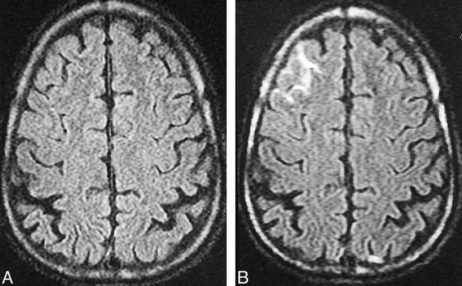
    fig. 1. A–E, Axial contrast-enhanced FLAIR images (A) (TR/TEeff/excitations = 10,002/158.8/1, TI = 2200; 5-mm-thick sections) show linear increased signal in several sulci over both convexities. Corresponding T1-weighted images (B) show no analogous abnormality. Coronal FLAIR image (C) (10,002/158.8/1, TI = 2200) obtained 6 hours after the axial image shows hyperintense CSF in sulci over both hemispheres (arrows). An unenhanced CT scan (D) was normal. Follow-up FLAIR image 2 days later (E) was also normal

  • Figure2
    • Download figure
    • Open in new tab
    • Download powerpoint

    fig. 2. A and B, Noncontrast FLAIR image (A) shows the expected dark cortical sulci over the frontal convexity. Contrast-enhanced FLAIR image (B) 15 minutes after injection of a triple dose of contrast material shows high signal in multiple sulci, most consistent with contrast effects

  • Figure3
    • Download figure
    • Open in new tab
    • Download powerpoint

    fig. 3. A and B, Sagittal FLAIR image (A) (10,007/189, TI = 2200; 5-mm-thick sections) in dog 2 immediately after injection of contrast agent (0.1 mmol/kg) reveals the expected low signal intensity of CSF in the fourth ventricle (arrow). Sagittal FLAIR image 30 minutes after contrast administration (B) shows that the CSF in the fourth ventricle is now isointense with surrounding brain (arrow)

  • Figure4
    • Download figure
    • Open in new tab
    • Download powerpoint

    fig. 4. A–C, Sagittal T1-weighted image (A) (550/15/1) obtained 6 hours after IV contrast administration (0.3 mmol/kg) shows the expected low-signal CSF in the cingulate sulcus (arrow). Sagittal FLAIR image (B), also obtained 6 hours after triple-dose contrast injection, shows abnormal high signal in the premedullary cistern (black arrow) as well as the cingulate sulcus (white arrow). The abnormal CSF signal on the coronal FLAIR image (C) is striking in the occipital sulci (arrows)

  • Figure5
    • Download figure
    • Open in new tab
    • Download powerpoint

    fig. 5. A and B, Images of the phantom obtained with FLAIR (10,000/154, TI = 1630) (A) and T1-weighted (400/16/1) (B) techniques. The lower tubes contain increasing dilutions of gadodiamide. There are more bright tubes in the lower two rows (lowest concentrations of contrast material) on the FLAIR image than on the T1-weighted image.

  • Figure6
    • Download figure
    • Open in new tab
    • Download powerpoint

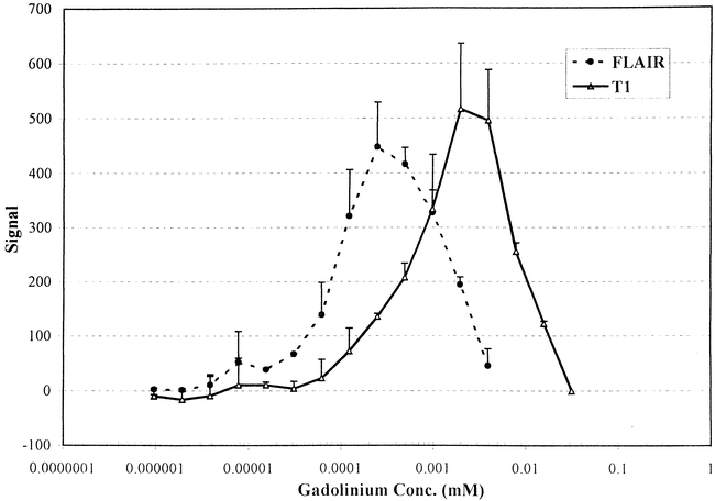
    fig. 6. Graph shows the effect of varying the concentrations of contrast material (range, 0.000001 to 0.5 mM) on the mean FLAIR and T1 signal intensity in the test tube phantom. The error bars represent +ISD and the data are from two separate experiments.

PreviousNext
Back to top

In this issue

American Journal of Neuroradiology
Vol. 21, Issue 1
1 Jan 2000
  • Table of Contents
  • Index by author
Advertisement
Print
Download PDF
Email Article

Thank you for your interest in spreading the word on American Journal of Neuroradiology.

NOTE: We only request your email address so that the person you are recommending the page to knows that you wanted them to see it, and that it is not junk mail. We do not capture any email address.

Enter multiple addresses on separate lines or separate them with commas.
Visualization of Intravenously Administered Contrast Material in the CSF on Fluid-AttenuatedInversion-Recovery MR Images: An In Vitro and Animal-Model Investigation
(Your Name) has sent you a message from American Journal of Neuroradiology
(Your Name) thought you would like to see the American Journal of Neuroradiology web site.
CAPTCHA
This question is for testing whether or not you are a human visitor and to prevent automated spam submissions.
Citation Tools
Visualization of Intravenously Administered Contrast Material in the CSF on Fluid-AttenuatedInversion-Recovery MR Images: An In Vitro and Animal-Model Investigation
Alexander C. Mamourian, P. Jack Hoopes, Lionel D. Lewis
American Journal of Neuroradiology Jan 2000, 21 (1) 105-111;

Citation Manager Formats

  • BibTeX
  • Bookends
  • EasyBib
  • EndNote (tagged)
  • EndNote 8 (xml)
  • Medlars
  • Mendeley
  • Papers
  • RefWorks Tagged
  • Ref Manager
  • RIS
  • Zotero
Share
Visualization of Intravenously Administered Contrast Material in the CSF on Fluid-AttenuatedInversion-Recovery MR Images: An In Vitro and Animal-Model Investigation
Alexander C. Mamourian, P. Jack Hoopes, Lionel D. Lewis
American Journal of Neuroradiology Jan 2000, 21 (1) 105-111;
del.icio.us logo Digg logo Reddit logo Twitter logo CiteULike logo Facebook logo Google logo Mendeley logo
  • Tweet Widget
  • Facebook Like
  • Google Plus One

Jump to section

  • Article
    • Abstract
    • Case Reports
    • Methods
    • Results
    • Discussion
    • Conclusion
    • Footnotes
    • References
  • Figures & Data
  • Info & Metrics
  • References
  • PDF

Related Articles

  • No related articles found.
  • PubMed
  • Google Scholar

Cited By...

  • Diagnosis and Prediction of Relapses in Susac Syndrome: A New Use for MR Postcontrast FLAIR Leptomeningeal Enhancement
  • HARMless: Transient Cortical and Sulcal Hyperintensity on Gadolinium-Enhanced FLAIR after Elective Endovascular Coiling of Intracranial Aneurysms
  • Pericortical Enhancement on Delayed Postgadolinium Fluid-Attenuated Inversion Recovery Images in Normal Aging, Mild Cognitive Impairment, and Alzheimer Disease
  • Presence of the hyperintense acute reperfusion marker on MRI after mechanical thrombectomy for large vessel occlusion is associated with worse early neurological recovery
  • Leptomeningeal gadolinium enhancement across the spectrum of chronic neuroinflammatory diseases
  • Gadolinium-based MRI characterization of leptomeningeal inflammation in multiple sclerosis
  • Blood-Brain Barrier Disruption after Cardiac Surgery
  • The Hyperintense Acute Reperfusion Marker on Fluid-Attenuated Inversion Recovery Magnetic Resonance Imaging Is Caused by Gadolinium in the Cerebrospinal Fluid
  • Comparison of the Added Value of Contrast-Enhanced 3D Fluid-Attenuated Inversion Recovery and Magnetization-Prepared Rapid Acquisition of Gradient Echo Sequences in Relation to Conventional Postcontrast T1-Weighted Images for the Evaluation of Leptomeningeal Diseases at 3T
  • Delayed CSF Enhancement in Posterior Reversible Encephalopathy Syndrome
  • Exceeding the Limits of the Normal Blood-Brain Barrier: Quo Vadis Gadolinium?
  • Increased Signal in the Subarachnoid Space on Fluid-Attenuated Inversion Recovery Imaging Associated with the Clearance Dynamics of Gadolinium Chelate: A Potential Diagnostic Pitfall
  • Transthyretin-Related Familial Amyloid Polyneuropathy: Evaluation of CSF Enhancement on Serial T1-Weighted and Fluid-Attenuated Inversion Recovery Images following Intravenous Contrast Administration
  • Intracranial Meningeal Disease: Comparison of Contrast-Enhanced MR Imaging with Fluid-Attenuated Inversion Recovery and Fat-Suppressed T1-Weighted Sequences
  • Perfusion MR Neuroimaging in Patients Undergoing Balloon Test Occlusion of the Internal Carotid Artery
  • Persistence of Gadolinium in CSF: A Diagnostic Pitfall in Patients with End-stage Renal Disease
  • Leptomeningeal Enhancement and Extravasation of Contrast Medium into the CSF Space? Response to the Preceding 2 Letters
  • Crossref
  • Google Scholar

This article has not yet been cited by articles in journals that are participating in Crossref Cited-by Linking.

More in this TOC Section

  • Usefulness of Quantitative Susceptibility Mapping for the Diagnosis of Parkinson Disease
  • White Matter Alterations in the Brains of Patients with Active, Remitted, and Cured Cushing Syndrome: A DTI Study
  • Qualitative and Quantitative Analysis of MR Imaging Findings in Patients with Middle Cerebral Artery Stroke Implanted with Mesenchymal Stem Cells
Show more BRAIN

Similar Articles

Advertisement

News and Updates

  • Lucien Levy Best Research Article Award
  • Thanks to our 2020 Distinguished Reviewers
  • Press Releases

Resources

  • Evidence-Based Medicine Level Guide
  • How to Participate in a Tweet Chat
  • AJNR Podcast Archive
  • Ideas for Publicizing Your Research
  • Librarian Resources
  • Terms and Conditions

Opportunities

  • Share Your Art in Perspectives
  • Get Peer Review Credit from Publons
  • Moderate a Tweet Chat

American Society of Neuroradiology

  • Neurographics
  • ASNR Annual Meeting
  • Fellowship Portal
  • Position Statements

© 2021 by the American Society of Neuroradiology | Print ISSN: 0195-6108 Online ISSN: 1936-959X

Powered by HighWire