
Article Figures & Data
Figures
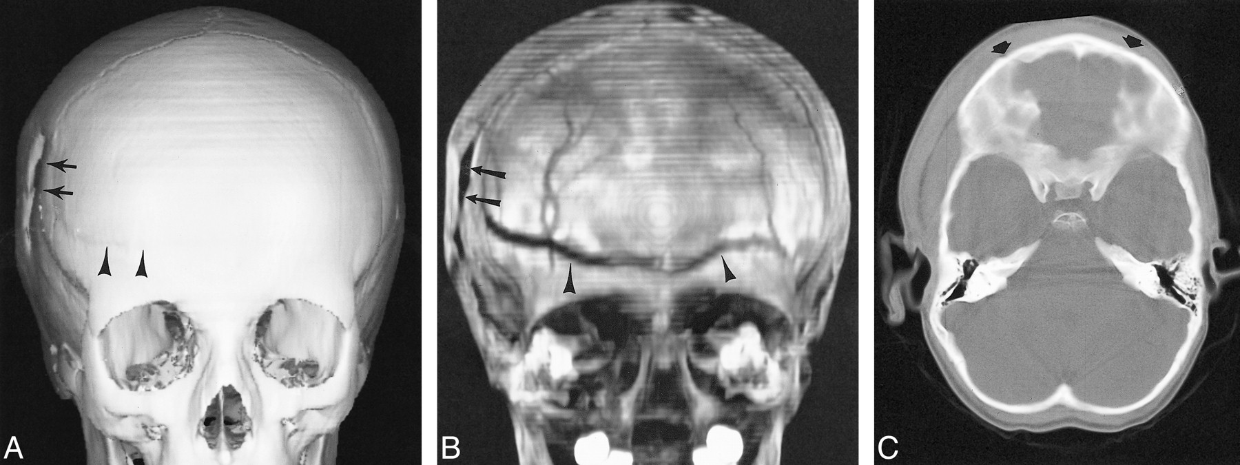
- fig 1.
Patient with Antley-Bixler syndrome, who is at risk of lambdoid craniosynostosis.>> A, Posterior projection of 3D CT SSD appears to show closure of the lambdoid suture (arrows).>>B, 2D axial CT image is equivocal in showing left lambdoid patency (arrow).>>C, Plain radiograph obtained the same day reveals an open right lambdoid suture (arrowheads) and questionable patent left lambdoid suture (small arrows).>>D, 3D CT MIP clearly demonstrates patent lambdoid suture bilaterally (small arrowheads)
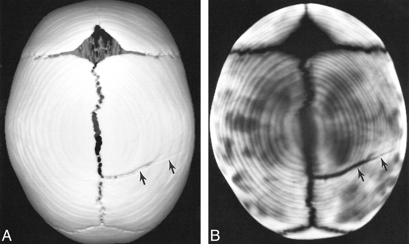
- fig 2.
Patient with sagittal craniosynostosis.>>A, Superior view of 3D CT SSD shows complete closure of the superior sagittal suture.>>B, Comparable superior view of 3D CT MIP characterizes the sagittal suture synostosis further by showing complete closure of the anterior and mid suture (arrows), but still incomplete closure posteriorly, with surrounding sclerosis (arrowheads)
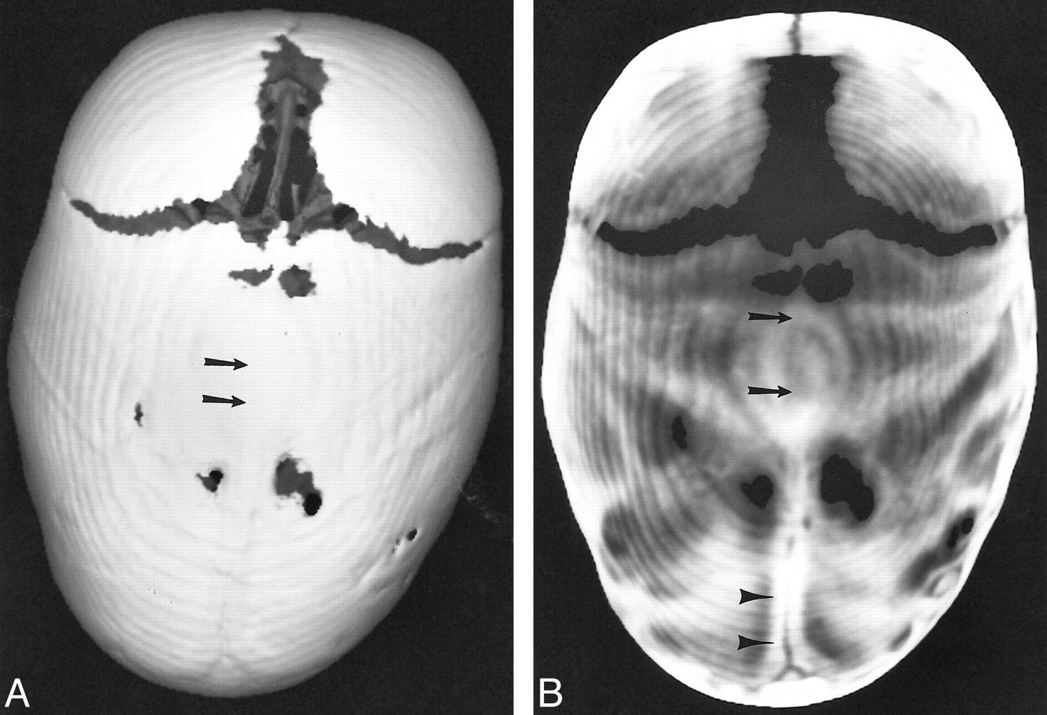
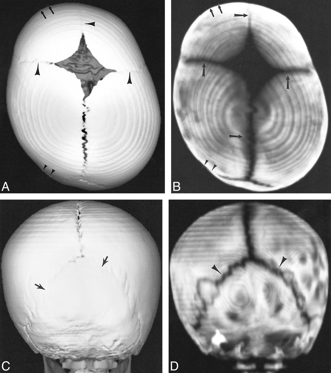
- fig 3.
Patient with positional molding.>>A, Superior projection of 3D CT SSD shows patent sagittal suture and probable patency of coronal and metopic sutures (arrowheads).>>B, Same projection with 3D CT MIP shows clearly patency of the coronal, sagittal, and metopic sutures (arrows). Both reconstructions show characteristic flattening of the posterior calvaria (small arrows) and ipsilateral frontal bone bossing (small arrowheads).>>C, Posterior projection of 3D CT SSD shows questionable patency of the entire lambdoid suture (arrows).>>D, Same projection with 3D CT MIP reveals unequivocal patency of lambdoid suture bilaterally (arrowheads)
- fig 4.
Patient with depressed right parietal fracture extending into the frontal bone.>>A, Frontal projection of 3D CT SSD reveals the depressed parietal fracture (arrows) but poorly outlines the extent of the frontal fracture (arrowheads).>>B, Comparable 3D CT MIP projection not only reveals the depressed parietal fracture (arrows) but the full extent of the frontal fracture (arrowheads).>>C, 2D axial CT failed to show the nondepressed frontal fracture component running parallel to sectioning plane (arrows)
- fig 5.
Patient with non-depressed parietal fracture.>>A, Superior projection of 3D CT SSD blends the margins of the fracture with the calvaria, making assessment of the extent of the fracture less conspicuous.>>B, Comparable 3D CT MIP shows the full extent of the fracture by separating the high density bone from the low-density fracture. Also note that conspicuity of all open sutures is well shown with 3D CT MIP


In this issue


Jump to section
Related Articles
Cited By...
- No citing articles found.