
Article Figures & Data
Figures
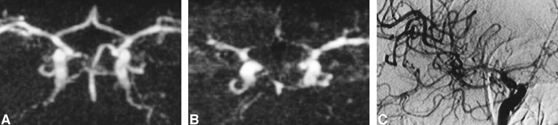
- fig 1.
A and B, MR angiograms (32/8/2; flip angle = 17°) obtained 1 (A) and 10 (B) days after acute SAH. At day 1, all vessels are normal (rating = 0); at day 10, a severe vasospasm is identified in both the ACA (rating = 3 bilaterally) and in the right MCA (rating = 3). In the left MCA, the narrowing is moderate (rating = 2) and a slight narrowing of both ICAs was also recorded (rating = 1 bilaterally) (not shown). The distal segments of arteries are poorly visualized, and the overall quality of MR angiography looks poor. These findings are typical of severe vasospasm and are related to the decreased flow into spastic vessels.
C, IA-DSA (right carotid arteriogram) obtained 9 days after SAH and 15 hours before MR angiography (B). Note presence of severe vasospasm (rating = 3 for the ACA and 2 for the MCA and ICA). Correlation with MR angiography was excellent for the ACA and good for the MCA and ICA.
- fig 2.
A, IA-DSA (right carotid arteriogram) obtained 8 days after SAH. An 8-mm aneurysm (arrowhead) is apparent in the right MCA and a slight vasospasm is seen in the MCA near the aneurysm (long arrows; rating = 1). The left ACA (A1 segment) is hypoplastic (short arrows).
B, MR angiogram (32/8/2; flip angle = 17°) obtained 3 hours before IA-DSA. The presence of methemoglobin (asterisk) decreases the image quality but interpretation is still possible. Only the distal segments beyond the right MCA trifurcation cannot be delineated owing to the superimposition of hyperintense signal arising from methemoglobin. The aneurysm is clearly identified (arrowhead) as well as the narrowing of the right MCA (long arrow; rating = 1) and the hypoplasia of the left ACA (short arrows). Correlation with IA-DSA appearance is excellent.
- fig 3.
A, IA-DSA (right carotid arteriogram) obtained 16 days after acute SAH. A 5-mm aneurysm is visible in the AComA (arrowheads). There is a slight narrowing of the ICA (rating = 1), but overall there is no angiographic vasospasm.
B and C, MR angiograms (32/8/2; flip angle = 17°) obtained 2 hours before IA-DSA. The aneurysm is clearly visible (arrowheads). On the MIP image (B), some portions of arteries are poorly visualized on both sides, suggesting the presence of a significant vasospasm (rating = 2 for both ICA and ACA and 1 for both MCAs). This is a false interpretation, which can be avoided by looking at the native slices (C). Significant pulsation artifacts (arrow) are present around the arteries, leading to a discontinuous aspect of vessels on the MIP image.
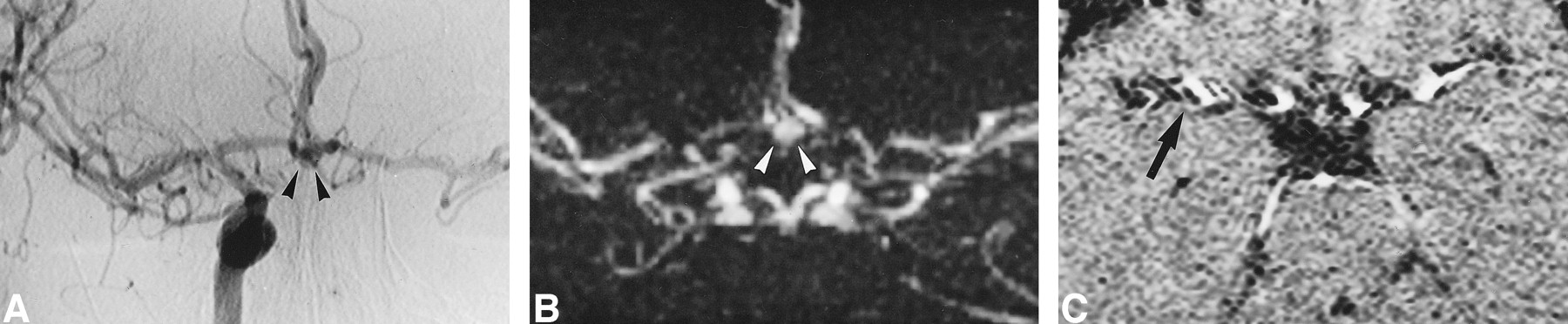
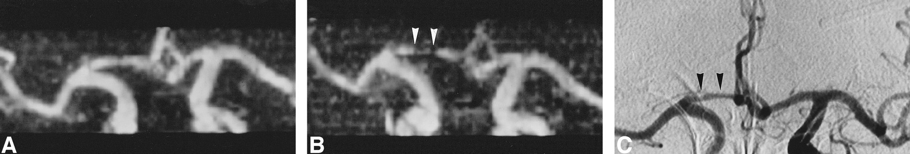
- fig 4.
A and B, MR angiograms (32/8/2; flip angle = 17°) obtained 2 (A) and 9 (B) days after acute SAH. At day 2, all vessels are normal (rating = 0); at day 9, significant vasospasm of the right ACA is identified (A1 segment, rating = 2) (arrowheads, B). The diagnosis is easy when a previous MR angiogram is available for comparison.
C, IA-DSA (right carotid arteriogram) obtained 10 days after SAH and 18 hours after MR angiography (B). The narrowing of the right A1 segment (arrowheads; rating = 2) was thought to be hypoplasia by one of the reviewers, but MR angiography performed before the spasm proved this interpretation to be incorrect.


Tables
 TABLE 1:
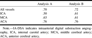
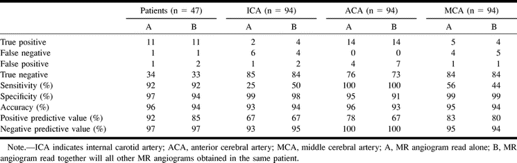
Weighted {κ} statistics showing the agreement between MR angiography and IA-DSA in detecting vasospasm
In this issue


Jump to section
Related Articles
Cited By...
- Assessment of Heating on Titanium Alloy Cerebral Aneurysm Clips during 7T MRI
- Evaluation of the JRecan device for thrombus retrieval: efficacy and safety in a swine model of acute arterial occlusion
- Neuroradiologic Diagnosis of Minor Leak prior to Major SAH: Diagnosis by T1-FLAIR Mismatch
- Early Intensive Versus Minimally Invasive Approach to Postoperative Hemodynamic Management After Subarachnoid Hemorrhage
- Experimental Evaluation of Immediate Recanalization Effect and Recanalization Efficacy of a New Thrombus Retriever for Acute Stroke Treatment In Vivo
- In Vivo Evaluation of the First Dedicated Combined Flow-Restoration and Mechanical Thrombectomy Device in a Swine Model of Acute Vessel Occlusion
- In Vivo Evaluation of the Phenox CRC Mechanical Thrombectomy Device in a Swine Model of Acute Vessel Occlusion
- Mechanical Thromboembolectomy for Acute Ischemic Stroke: Comparison of the Catch Thromboectomy Device and the Merci Retriever In Vivo
- Recurrent primary thunderclap headache and benign CNS angiopathy: Spectra of the same disorder?
- Mechanical Thrombectomy for Acute Ischemic Stroke: Thrombus-Device Interaction, Efficiency, and Complications In Vivo