Advertisement

Main menu

  • Home
  • Content
    • Current Issue
    • Publication Preview--Ahead of Print
    • Past Issue Archive
    • Case of the Week Archive
    • Classic Case Archive
    • Case of the Month Archive
  • For Authors
  • About Us
    • About AJNR
    • Editors
    • American Society of Neuroradiology
  • Submit a Manuscript
  • Podcasts
    • Subscribe on iTunes
    • Subscribe on Stitcher
  • More
    • Subscribers
    • Permissions
    • Advertisers
    • Alerts
    • Feedback
  • Other Publications
    • ajnr

User menu

  • Subscribe
  • Alerts
  • Log in

Search

  • Advanced search
American Journal of Neuroradiology

American Journal of Neuroradiology

  • Subscribe
  • Alerts
  • Log in

Advanced Search

  • Home
  • Content
    • Current Issue
    • Publication Preview--Ahead of Print
    • Past Issue Archive
    • Case of the Week Archive
    • Classic Case Archive
    • Case of the Month Archive
  • For Authors
  • About Us
    • About AJNR
    • Editors
    • American Society of Neuroradiology
  • Submit a Manuscript
  • Podcasts
    • Subscribe on iTunes
    • Subscribe on Stitcher
  • More
    • Subscribers
    • Permissions
    • Advertisers
    • Alerts
    • Feedback
  • Follow AJNR on Twitter
  • Visit AJNR on Facebook
  • Follow AJNR on Instagram
  • Join AJNR on LinkedIn
  • RSS Feeds
Research ArticleBRAIN

Magnetization Transfer Imaging and Proton MR Spectroscopy in the Evaluation of Axonal Injury: Correlation with Clinical Outcome after Traumatic Brain Injury

Grant Sinson, Linda J. Bagley, Kim M. Cecil, Maria Torchia, Joseph C. McGowan, Robert E. Lenkinski, Tracy K. McIntosh and Robert I. Grossman
American Journal of Neuroradiology January 2001, 22 (1) 143-151;
Grant Sinson
  • Find this author on Google Scholar
  • Find this author on PubMed
  • Search for this author on this site
Linda J. Bagley
  • Find this author on Google Scholar
  • Find this author on PubMed
  • Search for this author on this site
Kim M. Cecil
  • Find this author on Google Scholar
  • Find this author on PubMed
  • Search for this author on this site
Maria Torchia
  • Find this author on Google Scholar
  • Find this author on PubMed
  • Search for this author on this site
Joseph C. McGowan
  • Find this author on Google Scholar
  • Find this author on PubMed
  • Search for this author on this site
Robert E. Lenkinski
  • Find this author on Google Scholar
  • Find this author on PubMed
  • Search for this author on this site
Tracy K. McIntosh
  • Find this author on Google Scholar
  • Find this author on PubMed
  • Search for this author on this site
Robert I. Grossman
  • Find this author on Google Scholar
  • Find this author on PubMed
  • Search for this author on this site
  • Article
  • Figures & Data
  • Info & Metrics
  • References
  • PDF
Loading

Article Figures & Data

Figures

  • Tables
  • fig 1.
    • Download figure
    • Open in new tab
    • Download powerpoint
    fig 1.

    T2-weighted MR image (3000/90/1) at the level of the splenium. Asterisks indicate the regions of sampling for MTR acquisition; the box outlines the voxel for MR spectroscopy acquisition
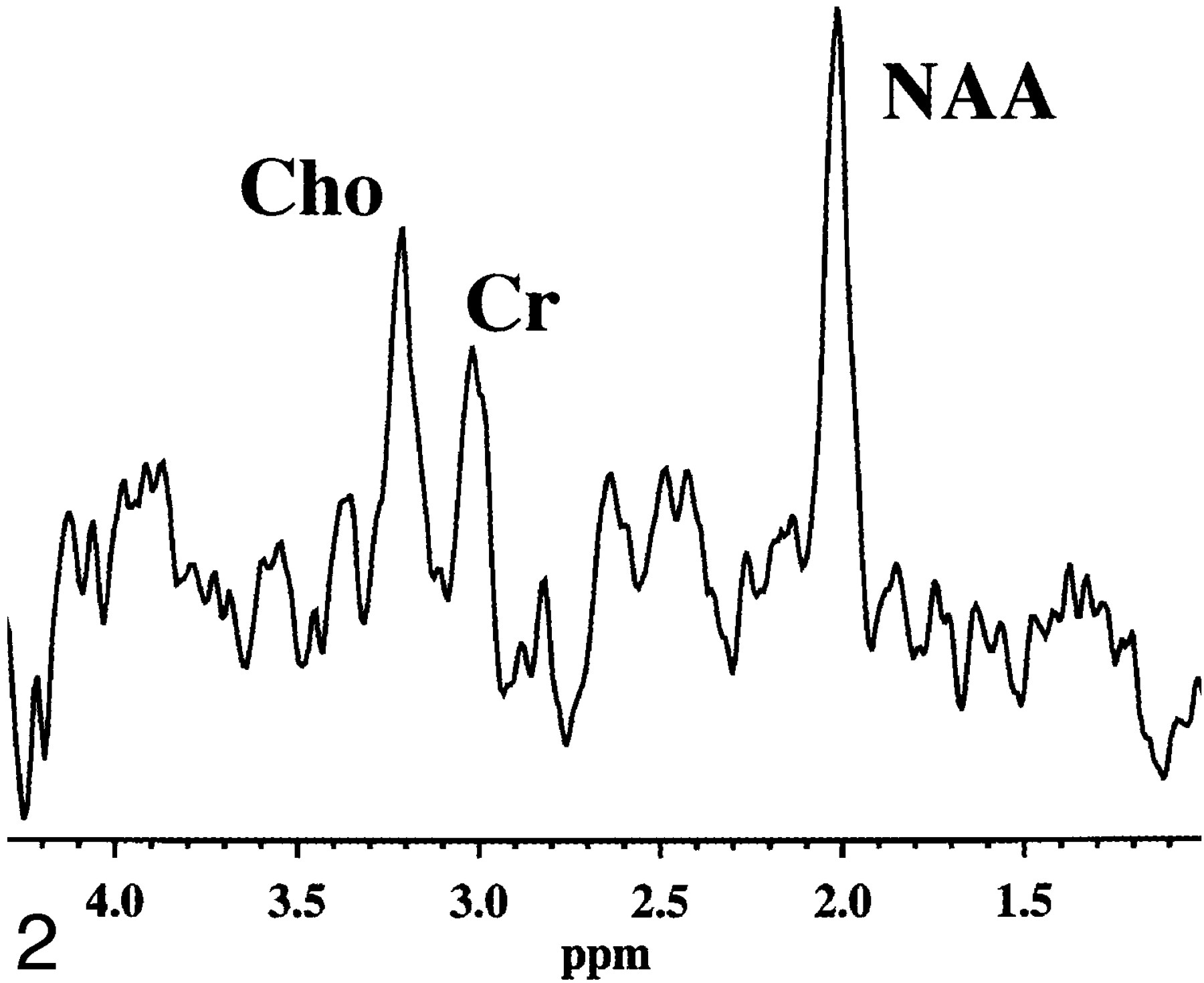
  • fig 2.
    • Download figure
    • Open in new tab
    • Download powerpoint
    fig 2.

    Proton MR spectrum obtained from normal splenium. The NAA/Cr ratio is 1.75. All spectra were acquired using a STEAM sequence (2000/31).
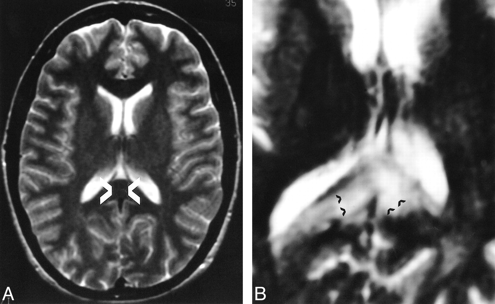
  • fig 3.
    • Download figure
    • Open in new tab
    • Download powerpoint
    fig 3.

    T2-weighted MR images (3000/90/1) at the level of the splenium in two different patients.

    A, Despite the normal appearance of the white matter in the splenium (arrows), this patient had an MTR greater than 2 SD below normal values.

    B, This patient has typical high signal in the region of the splenium (arrows), considered an indicator of severe diffuse injury.
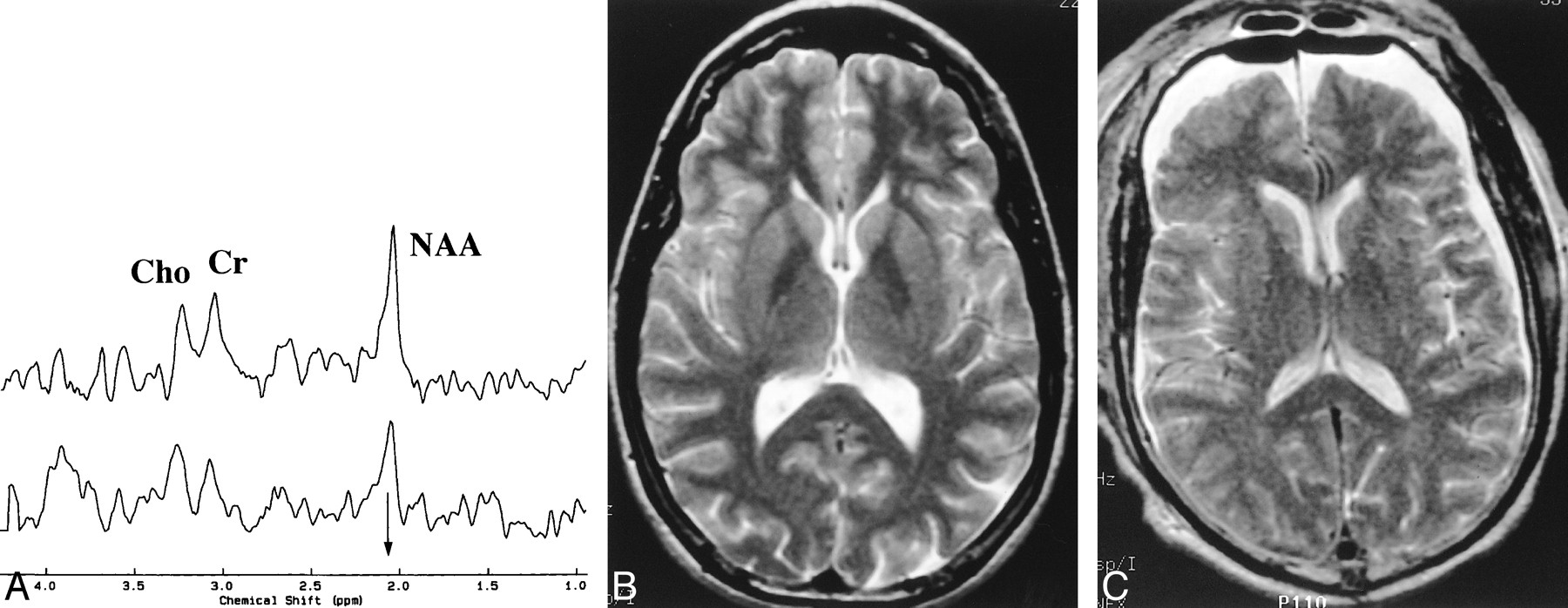
  • fig 4.
    • Download figure
    • Open in new tab
    • Download powerpoint
    fig 4.

    A, Proton MR spectra obtained from the splenium of two patients with head injury. The arrow identifies the methyl resonance of NAA, which is at 2.02 parts per million. The top spectrum was recorded in a patient with an initial GCS of 14 and a GOS of 5 and an NAA/Cr of 1.52. The bottom spectrum shows a lowering in the NAA peak that results in a decreased NAA/Cr ratio of 1.16 in a patient with a GCS of 4 and GOS of 2.

    B, T2-weighted MR image (2700/85/1) from patient in top spectrum in A.

    C, T2-weighted MR image (2700/80/1) from patient in bottom spectrum in A shows postoperative changes from evacuation of a subdural hematoma and bilateral hygromas.
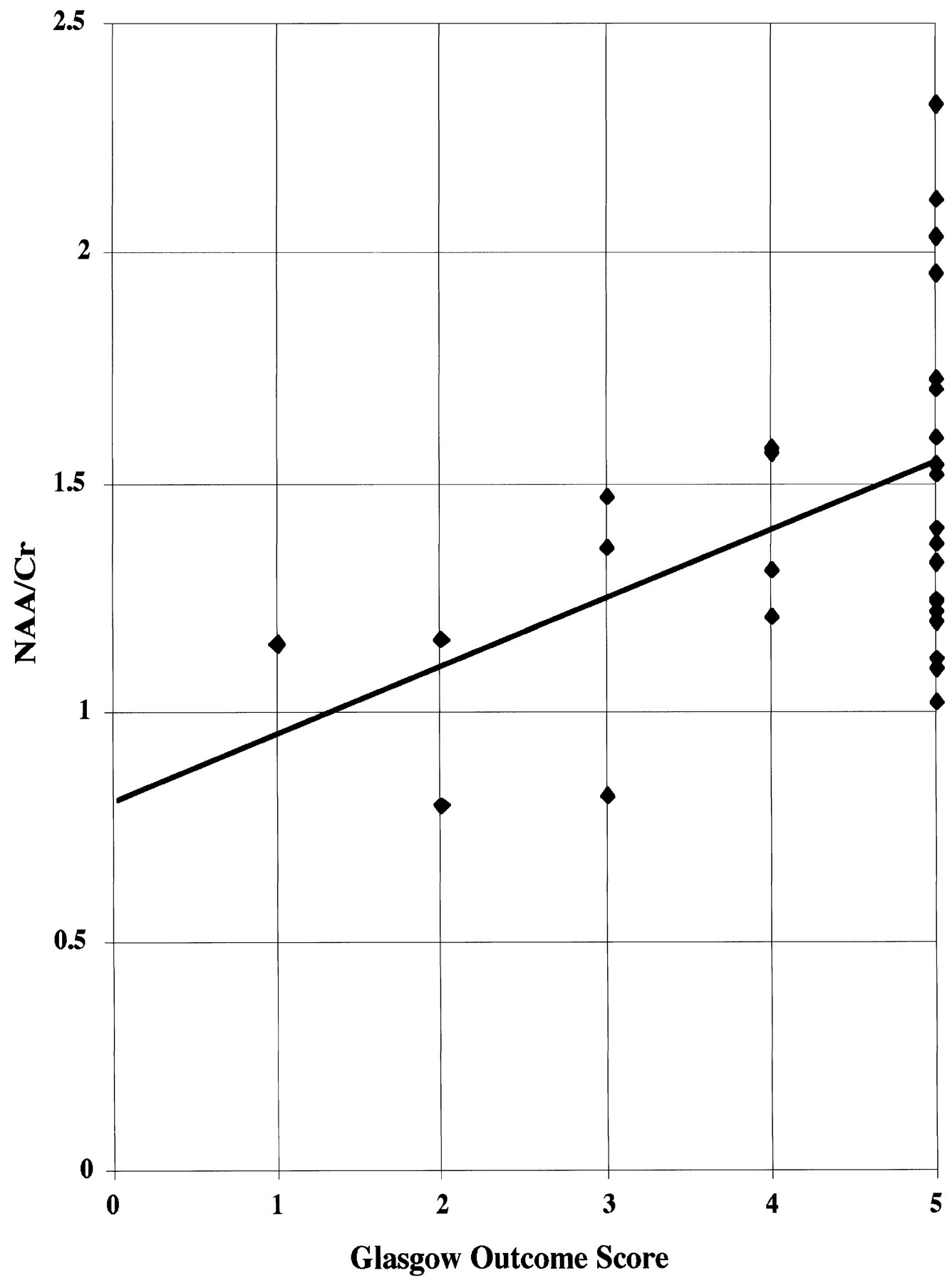
  • fig 5.
    • Download figure
    • Open in new tab
    • Download powerpoint
    fig 5.

    Relationship between NAA/Cr ratio and GOS score. Spearman's rank-order correlation indicates a significant correlation between NAA/Cr and outcome (Y = 0.142X + 0.819, rs = 0.4378, P < .01).
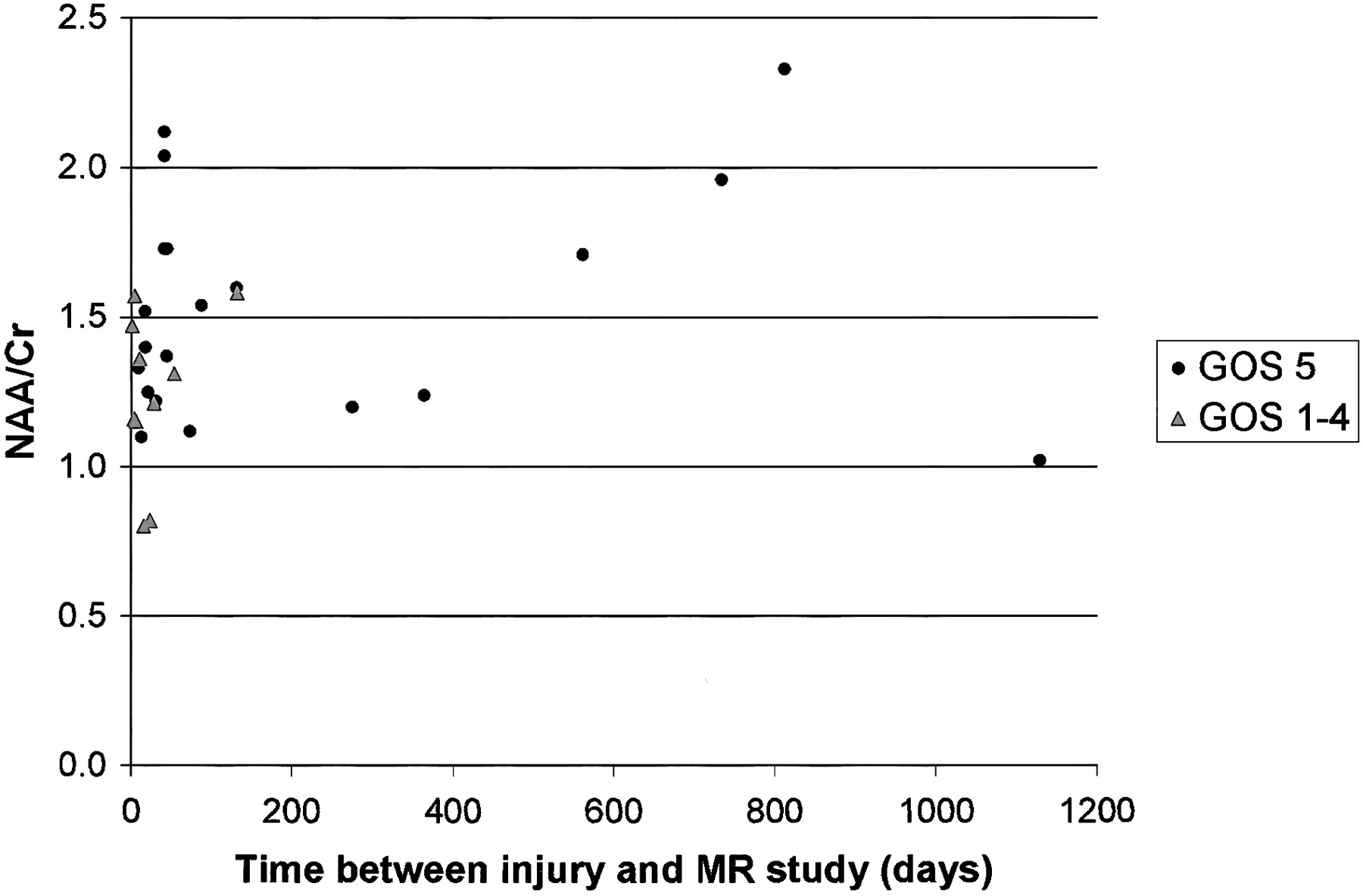
  • fig 6.
    • Download figure
    • Open in new tab
    • Download powerpoint
    fig 6.

    Relationship between NAA/Cr ratio and time interval from injury to imaging examination in both outcome groups. There was no significant correlation in either group.

Tables

  • Figures
  • Table1

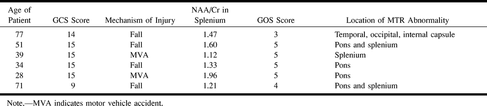
    Patients with abnormal MTR in NAWM

PreviousNext
Back to top

In this issue

American Journal of Neuroradiology
Vol. 22, Issue 1
1 Jan 2001
  • Table of Contents
  • Index by author
Advertisement
Print
Download PDF
Email Article

Thank you for your interest in spreading the word on American Journal of Neuroradiology.

NOTE: We only request your email address so that the person you are recommending the page to knows that you wanted them to see it, and that it is not junk mail. We do not capture any email address.

Enter multiple addresses on separate lines or separate them with commas.
Magnetization Transfer Imaging and Proton MR Spectroscopy in the Evaluation of Axonal Injury: Correlation with Clinical Outcome after Traumatic Brain Injury
(Your Name) has sent you a message from American Journal of Neuroradiology
(Your Name) thought you would like to see the American Journal of Neuroradiology web site.
CAPTCHA
This question is for testing whether or not you are a human visitor and to prevent automated spam submissions.
Citation Tools
Magnetization Transfer Imaging and Proton MR Spectroscopy in the Evaluation of Axonal Injury: Correlation with Clinical Outcome after Traumatic Brain Injury
Grant Sinson, Linda J. Bagley, Kim M. Cecil, Maria Torchia, Joseph C. McGowan, Robert E. Lenkinski, Tracy K. McIntosh, Robert I. Grossman
American Journal of Neuroradiology Jan 2001, 22 (1) 143-151;

Citation Manager Formats

  • BibTeX
  • Bookends
  • EasyBib
  • EndNote (tagged)
  • EndNote 8 (xml)
  • Medlars
  • Mendeley
  • Papers
  • RefWorks Tagged
  • Ref Manager
  • RIS
  • Zotero
Share
Magnetization Transfer Imaging and Proton MR Spectroscopy in the Evaluation of Axonal Injury: Correlation with Clinical Outcome after Traumatic Brain Injury
Grant Sinson, Linda J. Bagley, Kim M. Cecil, Maria Torchia, Joseph C. McGowan, Robert E. Lenkinski, Tracy K. McIntosh, Robert I. Grossman
American Journal of Neuroradiology Jan 2001, 22 (1) 143-151;
del.icio.us logo Digg logo Reddit logo Twitter logo CiteULike logo Facebook logo Google logo Mendeley logo
  • Tweet Widget
  • Facebook Like
  • Google Plus One

Jump to section

  • Article
    • Abstract
    • Methods
    • Results
    • Discussion
    • Conclusion
    • Footnotes
    • References
  • Figures & Data
  • Info & Metrics
  • References
  • PDF

Related Articles

  • No related articles found.
  • PubMed
  • Google Scholar

Cited By...

  • Pediatric Sports-Related Concussion Produces Cerebral Blood Flow Alterations
  • Evaluation of Delayed Neuronal and Axonal Damage Secondary to Moderate and Severe Traumatic Brain Injury Using Quantitative MR Imaging Techniques
  • Whole-Brain N-Acetylaspartate: A Marker of the Severity of Mild Head Trauma
  • Proton MR Spectroscopy and MRI-Volumetry in Mild Traumatic Brain Injury
  • 1H-MR Spectroscopy, Magnetization Transfer, and Diffusion-Weighted Imaging in Alcoholic and Nonalcoholic Patients with Cirrhosis with Hepatic Encephalopathy.
  • Proton MR Spectroscopic Imaging Depicts Diffuse Axonal Injury in Children with Traumatic Brain Injury
  • Late Proton MR Spectroscopy in Children after Traumatic Brain Injury: Correlation with Cognitive Outcomes
  • A review of structural magnetic resonance neuroimaging
  • Diffusion Tensor Imaging as Potential Biomarker of White Matter Injury in Diffuse Axonal Injury
  • Magnetization transfer imaging in focal epilepsy
  • Brain Atrophy in Mild or Moderate Traumatic Brain Injury: A Longitudinal Quantitative Analysis
  • Crossref
  • Google Scholar

This article has not yet been cited by articles in journals that are participating in Crossref Cited-by Linking.

More in this TOC Section

  • Evaluating the Effects of White Matter Multiple Sclerosis Lesions on the Volume Estimation of 6 Brain Tissue Segmentation Methods
  • Quiet PROPELLER MRI Techniques Match the Quality of Conventional PROPELLER Brain Imaging Techniques
  • Predictors of Reperfusion in Patients with Acute Ischemic Stroke
Show more BRAIN

Similar Articles

Advertisement

News and Updates

  • Lucien Levy Best Research Article Award
  • Thanks to our 2020 Distinguished Reviewers
  • Press Releases

Resources

  • Evidence-Based Medicine Level Guide
  • How to Participate in a Tweet Chat
  • AJNR Podcast Archive
  • Ideas for Publicizing Your Research
  • Librarian Resources
  • Terms and Conditions

Opportunities

  • Share Your Art in Perspectives
  • Get Peer Review Credit from Publons
  • Moderate a Tweet Chat

American Society of Neuroradiology

  • Neurographics
  • ASNR Annual Meeting
  • Fellowship Portal
  • Position Statements

© 2021 by the American Society of Neuroradiology | Print ISSN: 0195-6108 Online ISSN: 1936-959X

Powered by HighWire