Advertisement

Main menu

  • Home
  • Content
    • Current Issue
    • Publication Preview--Ahead of Print
    • Past Issue Archive
    • Case of the Week Archive
    • Classic Case Archive
    • Case of the Month Archive
  • For Authors
  • About Us
    • About AJNR
    • Editors
    • American Society of Neuroradiology
  • Submit a Manuscript
  • Podcasts
    • Subscribe on iTunes
    • Subscribe on Stitcher
  • More
    • Subscribers
    • Permissions
    • Advertisers
    • Alerts
    • Feedback
  • Other Publications
    • ajnr

User menu

  • Subscribe
  • Alerts
  • Log in

Search

  • Advanced search
American Journal of Neuroradiology

American Journal of Neuroradiology

  • Subscribe
  • Alerts
  • Log in

Advanced Search

  • Home
  • Content
    • Current Issue
    • Publication Preview--Ahead of Print
    • Past Issue Archive
    • Case of the Week Archive
    • Classic Case Archive
    • Case of the Month Archive
  • For Authors
  • About Us
    • About AJNR
    • Editors
    • American Society of Neuroradiology
  • Submit a Manuscript
  • Podcasts
    • Subscribe on iTunes
    • Subscribe on Stitcher
  • More
    • Subscribers
    • Permissions
    • Advertisers
    • Alerts
    • Feedback
  • Follow AJNR on Twitter
  • Visit AJNR on Facebook
  • Follow AJNR on Instagram
  • Join AJNR on LinkedIn
  • RSS Feeds
OtherHEAD AND NECK

Intracanalicular Optic Nerve Meningioma: A Serious Diagnostic Pitfall

Alan Jackson, Tufail Patankar and Roger D. Laitt
American Journal of Neuroradiology June 2003, 24 (6) 1167-1170;
Alan Jackson
  • Find this author on Google Scholar
  • Find this author on PubMed
  • Search for this author on this site
Tufail Patankar
  • Find this author on Google Scholar
  • Find this author on PubMed
  • Search for this author on this site
Roger D. Laitt
  • Find this author on Google Scholar
  • Find this author on PubMed
  • Search for this author on this site
  • Article
  • Figures & Data
  • Info & Metrics
  • References
  • PDF
Loading

Article Figures & Data

Figures

  • Tables
  • Fig 1.
    • Download figure
    • Open in new tab
    • Download powerpoint
    Fig 1.

    Case 2.

    A, CT image from case 2, showing a small soft tissue mass at the apex of the right orbit (arrow). This image is a prediagnostic scan obtained before the patient’s referral to our center. The nerve enlargement is subtle and is difficult to appreciate because of the relatively thick sections. This abnormality was identified only on retrospective review.

    B, T1-weighted MR image, reconstructed along the optic pathways, showing slight widening of the soft tissue of the optic nerve–optic nerve sheath complex at the orbital apex. The abnormality is still subtle and could easily be overlooked.

    C, Coronal T1-weighted MR image also showing subtle increase in the size of the soft tissue signal intensity within the right optic canal.

    D, Postcontrast T1-weighted MR image, reconstructed along the optic pathways, showing enhancement surrounding the optic nerve within the optic canal (large arrows). There is en-plaque growth along the walls of the sulcus chiasmaticus, giving a “rose thorn” appearance (small arrows).

  • Fig 2.
    • Download figure
    • Open in new tab
    • Download powerpoint
    Fig 2.

    Case 3.

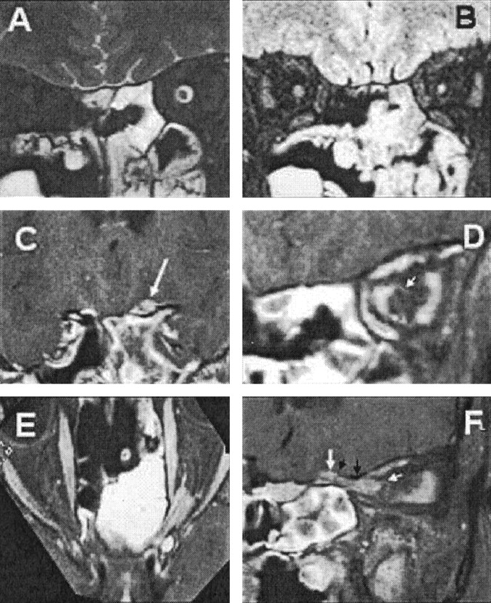
    A, T2-weighted coronal MR image, showing dilatation of the left optic nerve sheath.

    B, Heavily T2-weighted image with fat and water suppression (SPIR/FLAIR), showing increased T2 signal intensity and atrophy of the optic nerve.

    C, Coronal images through the sulcus chiasmaticus, demonstrating a small tumor nodule arising from the cranial end of the left optic canal (arrow).

    D, Coronal image from a contrast-enhanced T1-weighted volume-rendered MR image, acquired with fat suppression through the optic nerve in the midorbit, showing en-plaque growth of tumor within the left optic nerve sheath (arrow).

    E, Axial oblique reconstruction from a contrast-enhanced T1-weighted volume-rendered MR image, acquired with fat suppression along the optic canals, showing the extent of the tumor. Note the optic nerve emerging from the tumor anteriorly and the en-plaque growth of tumor along the anterior optic nerve sheath.

    F, Sagittal oblique reconstruction from a contrast-enhanced T1-weighted volume-rendered MR image, acquired with fat suppression along the course of the left optic canal, showing the extent of the tumor. Note the tram track sign due to the central nonenhancing optic nerve, the mural nodule (large white arrow) arising behind the roof of the optic canal (black arrows), and the en-plaque growth of the tumor along the anterior part of the orbital optic nerve sheath (small white arrow).

Tables

  • Figures
  • Clinical Data for Patients with Cannalicular Optic Nerve Meningloma

    PatientAge at Presentation (years)Time to Diagnosis (months)Surgical OutcomeOutcome
    12449No surgeryBlind
    22822CRVision preserved
    32832PRComplete loss of vision in right eye
    43321PRLight perception in left eye only
    53612CRVision preserved
    63814CRVision preserved
    • Note.—CR, complete resection; PR, partial resection.

PreviousNext
Back to top

In this issue

American Journal of Neuroradiology: 24 (6)
American Journal of Neuroradiology
Vol. 24, Issue 6
1 Jun 2003
  • Table of Contents
  • Index by author
Advertisement
Print
Download PDF
Email Article

Thank you for your interest in spreading the word on American Journal of Neuroradiology.

NOTE: We only request your email address so that the person you are recommending the page to knows that you wanted them to see it, and that it is not junk mail. We do not capture any email address.

Enter multiple addresses on separate lines or separate them with commas.
Intracanalicular Optic Nerve Meningioma: A Serious Diagnostic Pitfall
(Your Name) has sent you a message from American Journal of Neuroradiology
(Your Name) thought you would like to see the American Journal of Neuroradiology web site.
CAPTCHA
This question is for testing whether or not you are a human visitor and to prevent automated spam submissions.
Citation Tools
Intracanalicular Optic Nerve Meningioma: A Serious Diagnostic Pitfall
Alan Jackson, Tufail Patankar, Roger D. Laitt
American Journal of Neuroradiology Jun 2003, 24 (6) 1167-1170;

Citation Manager Formats

  • BibTeX
  • Bookends
  • EasyBib
  • EndNote (tagged)
  • EndNote 8 (xml)
  • Medlars
  • Mendeley
  • Papers
  • RefWorks Tagged
  • Ref Manager
  • RIS
  • Zotero
Share
Intracanalicular Optic Nerve Meningioma: A Serious Diagnostic Pitfall
Alan Jackson, Tufail Patankar, Roger D. Laitt
American Journal of Neuroradiology Jun 2003, 24 (6) 1167-1170;
del.icio.us logo Digg logo Reddit logo Twitter logo Facebook logo Google logo Mendeley logo
  • Tweet Widget
  • Facebook Like
  • Google Plus One

Jump to section

  • Article
    • Abstract
    • Case Reports and Results
    • Discussion
    • Conclusion
    • References
  • Figures & Data
  • Info & Metrics
  • References
  • PDF

Related Articles

  • No related articles found.
  • PubMed
  • Google Scholar

Cited By...

  • No citing articles found.
  • Crossref
  • Google Scholar

This article has not yet been cited by articles in journals that are participating in Crossref Cited-by Linking.

More in this TOC Section

  • Correlation of Apparent Diffusion Coefficient at 3T with Prognostic Parameters of Retinoblastoma
  • Parathyroid Lesions: Characterization with Dual-Phase Arterial and Venous Enhanced CT of the Neck
  • MR Diagnosis of Facial Neuritis: Diagnostic Performance of Contrast-Enhanced 3D-FLAIR Technique Compared with Contrast-Enhanced 3D-T1-Fast-Field Echo with Fat Suppression
Show more HEAD AND NECK

Similar Articles

Advertisement

News and Updates

  • Lucien Levy Best Research Article Award
  • Thanks to our 2021 Distinguished Reviewers
  • Press Releases

Resources

  • Evidence-Based Medicine Level Guide
  • How to Participate in a Tweet Chat
  • AJNR Podcast Archive
  • Ideas for Publicizing Your Research
  • Librarian Resources
  • Terms and Conditions

Opportunities

  • Share Your Art in Perspectives
  • Get Peer Review Credit from Publons
  • Moderate a Tweet Chat

American Society of Neuroradiology

  • Neurographics
  • ASNR Annual Meeting
  • Fellowship Portal
  • Position Statements

© 2022 by the American Society of Neuroradiology | Print ISSN: 0195-6108 Online ISSN: 1936-959X

Powered by HighWire