
Article Figures & Data
Figures
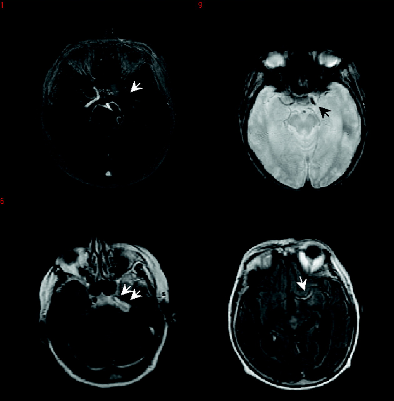
- Fig 1.
An 82-year-old female patient with cardioembolic stroke, baseline NIHSSS of 18 received rt-PA 88 minutes after symptom onset. Stroke MR imaging at 40 minutes after symptom onset. MRA (upper left) shows left-sided ICA occlusion extending into the MCA (arrows). This occlusion is seen on GRE (upper right, arrows) as a GRE SVS only at the level of the MCA, a FLAIR HVS can be seen at the level of the ICA and MCA (lower row, arrows).
Tables
PWI/MRA: Vessel Occlusion, Baseline CT baseline HMCAS FLAIR baseline HVS GRE baseline SVS No Yes Total No Yes Total No Yes Total No 6 0 6 9 3 12 9 3 12 Yes 15 10 25 15 29 44 29 15 44 Total 21 10 31 24 32 56 38 18 56 Sensitivity (%) 40 65.9 34.1 Specificity (%) 100 75 75 PPV (%) 100 90.6 83.3 NPV (%) 28.6 37.5 23.7 Accuracy (%) 51.6 67.9 42.9 PWI/MRA: Vessel Occlusion, 2 Hours FLAIR 2-Hour HVS GRE 2-Hour SVS No Yes Total No Yes Total No 20 0 20 14 6 20 Yes 11 25 36 18 18 36 Total 31 25 56 32 24 56 Sensitivity (%) 69.4 50 Specificity (%) 100 70 PPV (%) 100 75 NPV (%) 64.5 43.8 Accuracy (%) 80.4 57.1 PWI/MRA: Vessel Occlusion, 24 Hours FLAIR 24-Hour HVS GRE 24-Hour SVS No Yes Total No Yes Total No 29 1 30 26 4 30 Yes 13 13 26 12 14 26 Total 42 14 56 38 18 56 Sensitivity (%) 50 53.8 Specificity (%) 96.7 86.7 PPV (%) 92.9 77.8 NPV (%) 69 68.4 Accuracy (%) 75 71.4
In this issue


Jump to section
Related Articles
Cited By...
- FLAIR Vascular Hyperintensities as a Surrogate of Collaterals in Acute Stroke: DWI Matters
- SWI Susceptibility Vessel Sign in Patients Undergoing Mechanical Thrombectomy for Acute Ischemic Stroke
- Radiology-Pathology Correlations of Intracranial Clots: Current Theories, Clinical Applications, and Future Directions
- Clinical prognosis of FLAIR hyperintense arteries in ischaemic stroke patients: a systematic review and meta-analysis
- Erythrocyte Fraction Within Retrieved Thrombi Contributes to Thrombolytic Response in Acute Ischemic Stroke
- Do Fluid-Attenuated Inversion Recovery Vascular Hyperintensities Represent Good Collaterals before Reperfusion Therapy?
- Correlation of imaging and histopathology of thrombi in acute ischemic stroke with etiology and outcome: a systematic review
- Fluid-Attenuated Inversion Recovery Vascular Hyperintensity Topography, Novel Imaging Marker for Revascularization in Middle Cerebral Artery Occlusion
- Different risk factors for poor outcome between patients with positive and negative susceptibility vessel sign
- Fluid-Attenuated Inversion Recovery Vascular Hyperintensities-Diffusion-Weighted Imaging Mismatch Identifies Acute Stroke Patients Most Likely to Benefit From Recanalization
- Hyperintense Vessels on T2-PROPELLER-FLAIR in Patients with Acute MCA Stroke: Prediction of Arterial Stenosis and Perfusion Abnormality
- Significance of Development and Reversion of Collaterals on MRI in Early Neurologic Improvement and Long-Term Functional Outcome after Intravenous Thrombolysis for Ischemic Stroke
- Do FLAIR Vascular Hyperintensities beyond the DWI Lesion Represent the Ischemic Penumbra?
- Sensitivity and Specificity of the Hyperdense Artery Sign for Arterial Obstruction in Acute Ischemic Stroke
- Morphology of Susceptibility Vessel Sign Predicts Middle Cerebral Artery Recanalization After Intravenous Thrombolysis
- Hyperintense Basilar Artery on FLAIR MR Imaging: Diagnostic Accuracy and Clinical Impact in Patients with Acute Brain Stem Stroke
- Acute Stroke Imaging Research Roadmap II
- Clot Burden Score on Admission T2*-MRI Predicts Recanalization in Acute Stroke
- Clinical Significance of Fluid-Attenuated Inversion Recovery Vascular Hyperintensities in Transient Ischemic Attack
- Characterization of Arterial Thrombus Composition by Magnetic Resonance Imaging in a Swine Stroke Model
- Location of the Clot and Outcome of Perfusion Defects in Acute Anterior Circulation Stroke Treated with Intravenous Thrombolysis
- Hyperintense Vessels on Acute Stroke Fluid-Attenuated Inversion Recovery Imaging: Associations With Clinical and Other MRI Findings
- Use of neuroimaging to guide the treatment of patients beyond the 8-hour time window
- Hyperintense Vessel Sign on Fluid-Attenuated Inversion Recovery MR Imaging Is Reduced by Gadolinium
- Thrombus Branching and Vessel Curvature Are Important Determinants of Middle Cerebral Artery Trunk Recanalization With Merci Thrombectomy Devices
- Fluid-Attenuated Inversion Recovery Images and Stroke Outcome After Thrombolysis
- Sulcal Effacement on Fluid Attenuation Inversion Recovery Magnetic Resonance Imaging in Hyperacute Stroke: Association With Collateral Flow and Clinical Outcomes
- Fluid-Attenuated Inversion Recovery Vascular Hyperintensities: An Important Imaging Marker for Cerebrovascular Disease
- M1 Susceptibility Vessel Sign on T2* as a Strong Predictor for No Early Recanalization After IV-t-PA in Acute Ischemic Stroke
- Outcomes of Intravenous Recombinant Tissue Plasminogen Activator Therapy According to Gender: A Clinical Registry Study and Systematic Review
- Acute Ischemic Infarction Defined by a Region of Multiple Hypointense Vessels on Gradient-Echo T2* MR Imaging at 3T
- Distal hyperintense vessels on FLAIR: An MRI marker for collateral circulation in acute stroke?
- Angiography Reveals That Fluid-Attenuated Inversion Recovery Vascular Hyperintensities Are Due to Slow Flow, Not Thrombus
- Cardiogenic and Aortogenic Brain Embolism
- Recanalization after thrombolysis in stroke patients: Predictors and prognostic implications
- Temporal Profile of Recanalization After Intravenous Tissue Plasminogen Activator: Selecting Patients for Rescue Reperfusion Techniques
- Significance of Susceptibility Vessel Sign on T2*-Weighted Gradient Echo Imaging for Identification of Stroke Subtypes
- Imaging the Clot: Does Clot Appearance Predict the Efficacy of Thrombolysis?