
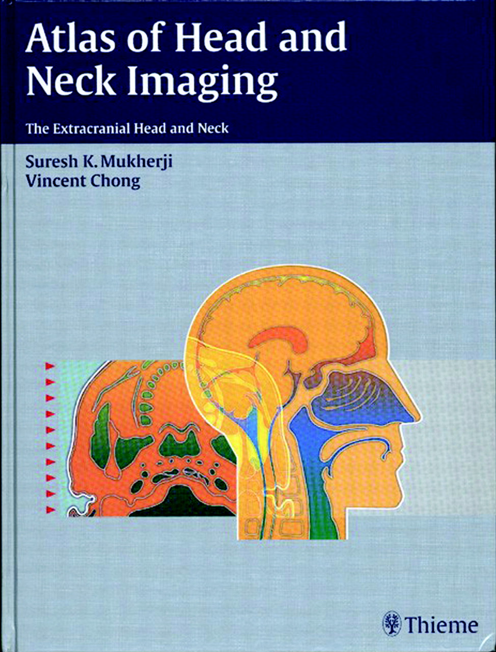
Suresh K. Mukherji and Vincent Chong. New York: Thieme; 2004. 602 pages, 811 illustrations, $169.
The number of teaching atlases in neuroradiology abound, and their popularity, in lieu of standard textbooks, seems to be increasing. The usefulness of such publications depend on the reader’s experience; for those involved with a large daily volume of neuroimaging where there is significant clinical feedback, these atlases are of less value than in situations where such volume and clinical correlation are not available. Each such atlas needs to be judged according to whether it covers all vital areas, demonstrates high-quality state-of-the-art images, and imparts complete imaging information. With these criteria in mind, Atlas of Head and Neck Imaging, by Suresh Mukherji and Vincent Chong, was reviewed.
This is a large book with clear, unambiguous descriptions of the most important diagnoses in head and neck radiology. Because of the way the book is organized, each diagnostic entity is called a different chapter, so what one ends up with is a book of 215 “chapters.” Actually the book is divided into 10 chapters, which are called sections, and within each section anywhere between six and 74 cases are presented. The sections are divided into more or less a standard way of dealing with disease in different areas, with the following covered: masticator space, parotid space, visceral space (which includes 74 cases), retropharyngeal space, prevertebral space (with six cases), parapharyngeal space, carotid space, buccal space, sublingual space, and submandibular space. The reader may be surprised that the book does not contain material on the temporal bone, orbits, or sinuses, but the authors made it clear that their intent was to concentrate on the extracranial head and neck.
The manner in which the book is laid out is pleasing. The outer portion of the book has gray tabs so one can quickly go from one section (i.e., one space) to another with a flick of the thumb. Although, at the beginning of each section there is a brief one-page summary of that space with line drawings, anatomic sections, and representative images (CT and MR) of the space under consideration, a reader is advised to have nearby a text that describes far more fully the normal anatomy. This will make the material in this text more understandable. Because there are so many structures in any give image, there could have been an overlabeling of each image that would have cluttered the material; fortunately, the authors avoided this and have stuck to labeling just the salient features. Because each chapter (i.e., case) is allotted a great deal of space with large margins and unused blank areas at the end of each case, the book is inordinately heavy. This reviewer was surprised to see such a large volume of unused space. In many chapters these blank areas could have been diminished by making the figures larger. Nonetheless, two advantages of this are that there is plenty of space to scribble notes relative to each case and one also becomes a “stronger” radiologist after reading the book and transporting it from place to place.
The appeal of this textbook would be for a radiologist with some experience in head and neck radiology and who has a reasonable understanding of the complex anatomy in this area. It certainly is not a primary text for those starting out to learn the imaging of this region. For those who want that sort of material, or in fact for any radiologist wanting a far more complete description of this area, the text by Som and Curtin is far more desirable. But in fairness, this book was written to be simply an atlas.
Each case has seven paragraphs with the following subheadings: epidemiology, clinical findings, pathology, treatment, imaging findings, imaging peaks, and suggested reading. For a text whose primary audience is radiologists, it is not clear why there is not also a section on differential diagnosis. Although some of the entities shown may have a limited, or perhaps no differential diagnosis, that is not the situation for most of the cases. For a radiologist, that perhaps would be one of the key elements he or she would be looking for in this type of atlas. Such a paragraph should have been labeled “major differential considerations,” limited to three or four alternatives. Although under some of the “imaging pearls” a differential diagnosis is mentioned, this is not true for many of the cases.
With 215 different cases shown, the atlas covers most every diagnosis one is liable to encounter in the extracranial head and neck. So the question is how do you use this book? Because it is not set up in a query-like manner, blinding yourself to the diagnosis is difficult. Similarly it is not set up to be read as a textbook, taking you from a complete description of normal imaging anatomy through the pathology as seen by CT and MR imaging. Probably the best way to use this atlas is to first look at the images and decide whether you are (or are not) familiar with that disease. If not, the outlined text is available for review.
Although this is not a text, most neuroradiologists would want to purchase it (but at $169.00, it is expensive for what you get). It will have particular appeal to those whose primary interest is in otorhynolaryngologic radiology, because it could be a helpful reference, and it also has a place in the library of any academic department. Thus, under those limited circumstances, it is recommended.
- Copyright © American Society of Neuroradiology
In this issue


Jump to section
Related Articles
Cited By...
- No citing articles found.