
Abstract
SUMMARY: We report an extremely rare case of a congenital spinal cord hamartoma in a male neonate who presented with upper extremity weakness and a port wine stain on the right upper extremity and chest. MR imaging findings are described, and the importance of localizing the lesion in the spinal canal with respect to the dura and its impact on neurosurgical management is stressed.
The presence of a congenital spinal mass requires adequate imaging with appropriate management. The differential diagnosis for these lesions includes teratomas, spinal dysraphism, lipomas, epidermoids, and dermoids.1 Another entity is the so-called spinal hamartoma, which is a very rare spinal tumor known to occur in children.2
Case Report
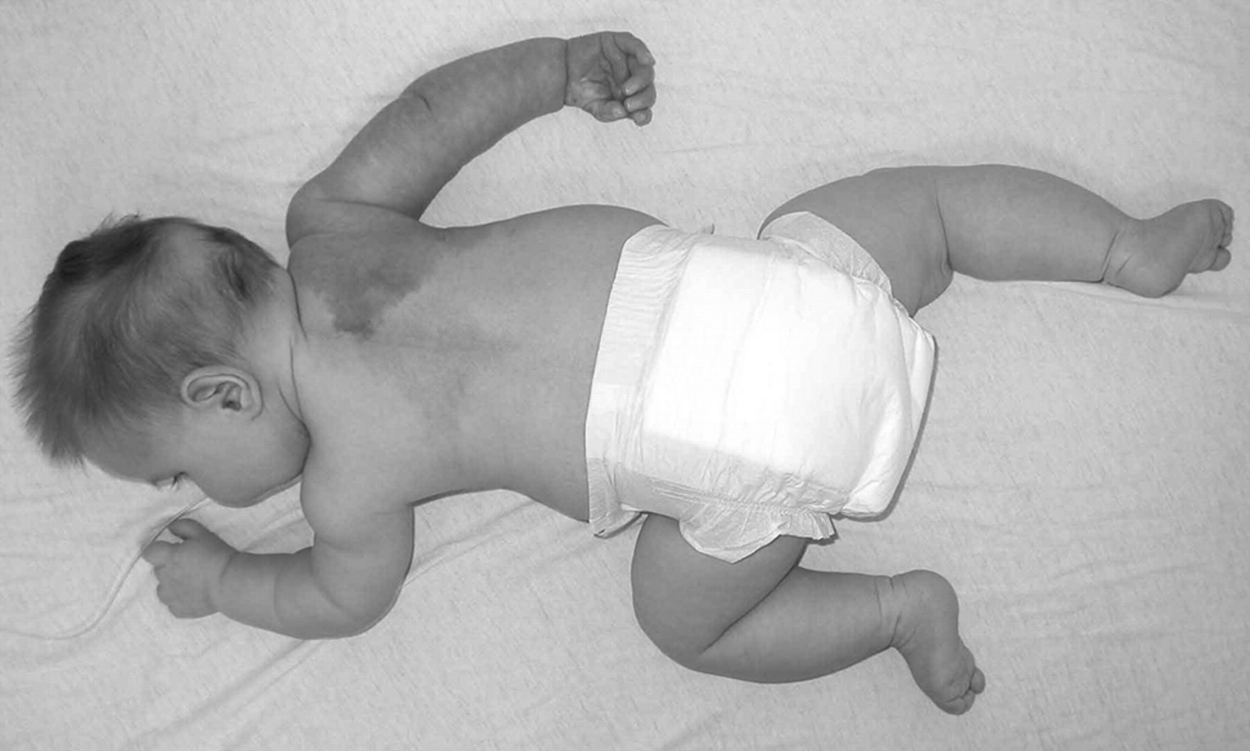
A neonate male, born by an uncomplicated caesarian delivery, presented with upper extremity hypotonia and decreased movement in the left upper extremity and little to no movement in the right upper extremity. A large port wine stain was noted on the right arm, upper thorax, and back (Fig 1).

Large port wine stain of the right arm, upper thorax, and back.
Initial MR imaging performed at 7 days of age in an outside institution demonstrated a large enhancing heterogenous cervicothoracic intraspinal tumor with extension to the right paravertebral and brachial plexus region through the right C7 and T1 neural foramen (Fig 2). The differential diagnosis entertained at this point was a nerve sheath tumor (based on the foraminal extension) or a teratoma. The patient was then referred to our institution for neurosurgical consultation. Resection of the intraspinal portion of the tumor was recommended with the objective of decompressing the cervical spinal canal, with a plan to deal with the paravertebral extension of the lesion at a later date. A C4 through T4 laminotomy was performed. The tumor was immediately identified within the spinal canal in the epidural space and was resected. The dura was not opened because this was assumed to be an extradural tumor based on the preoperative imaging and intraoperative findings. Pathologic findings demonstrated a very unusual lesion with well-formed elements usually found in the spinal and paraspinal region, including vessels, cartilage, nerves, and ganglion cells, which were all arranged in a helter-skelter fashion, consistent with a rare entity—spinal hamartoma (Figs 3 and 4).

A, Coronal T2-weighted MR image and (B) axial T2-weighted MR image demonstrate a large cervicothoracic intraspinal tumor with extension to the right paravertebral and brachial plexus region through the right C7 and T1 neural foramen. Tumor shows increased signal intensity on T2-weighted sequences (A, B). The cord is compressed and displaced laterally to the left by the mass (arrow).
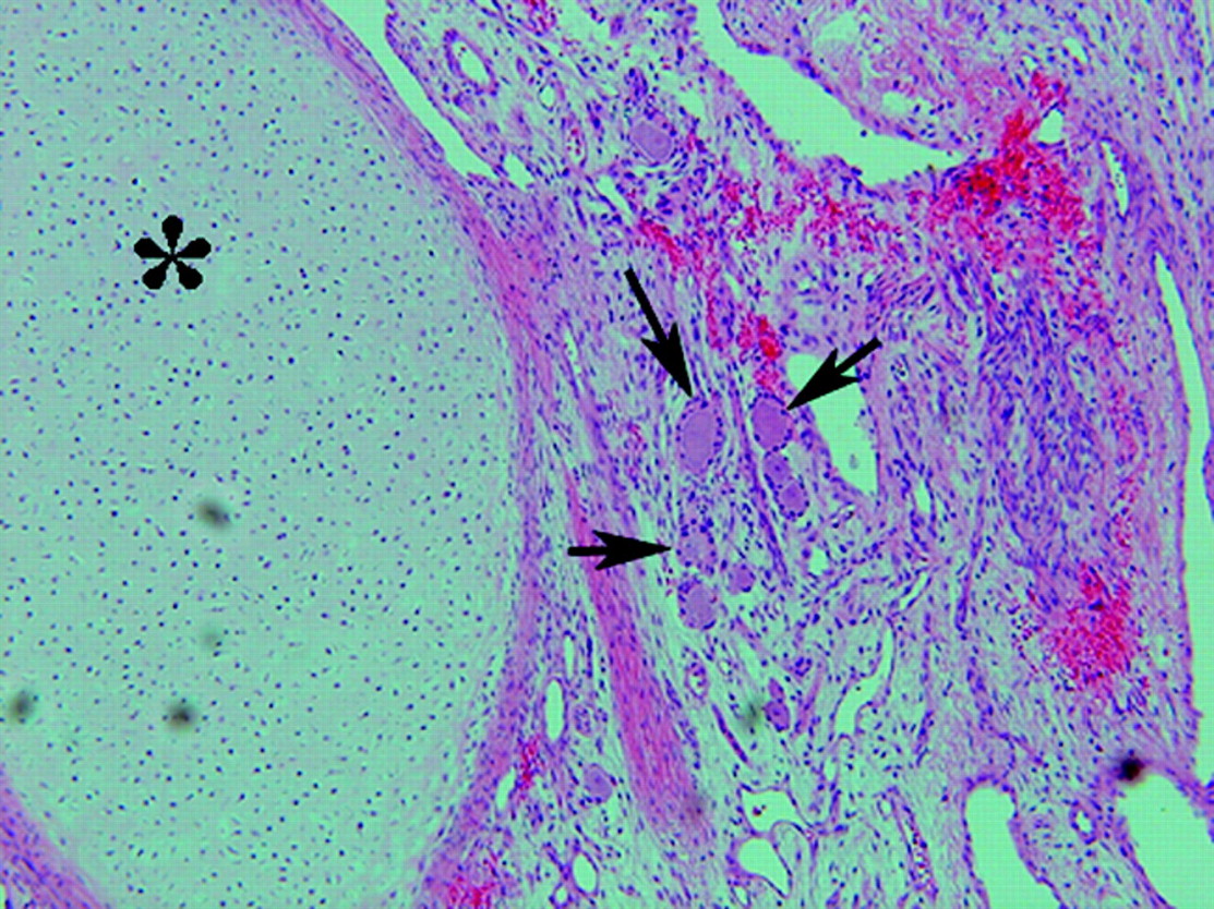
Photomicrograph shows histologic appearance of the lesion and demonstrates the presence of mature cartilage (asterisk) against a background of small- and intermediate-sized vessels, some containing smooth muscle in their walls. Ganglion cells are seen in the center (arrows) (H & E, original magnification ×100).
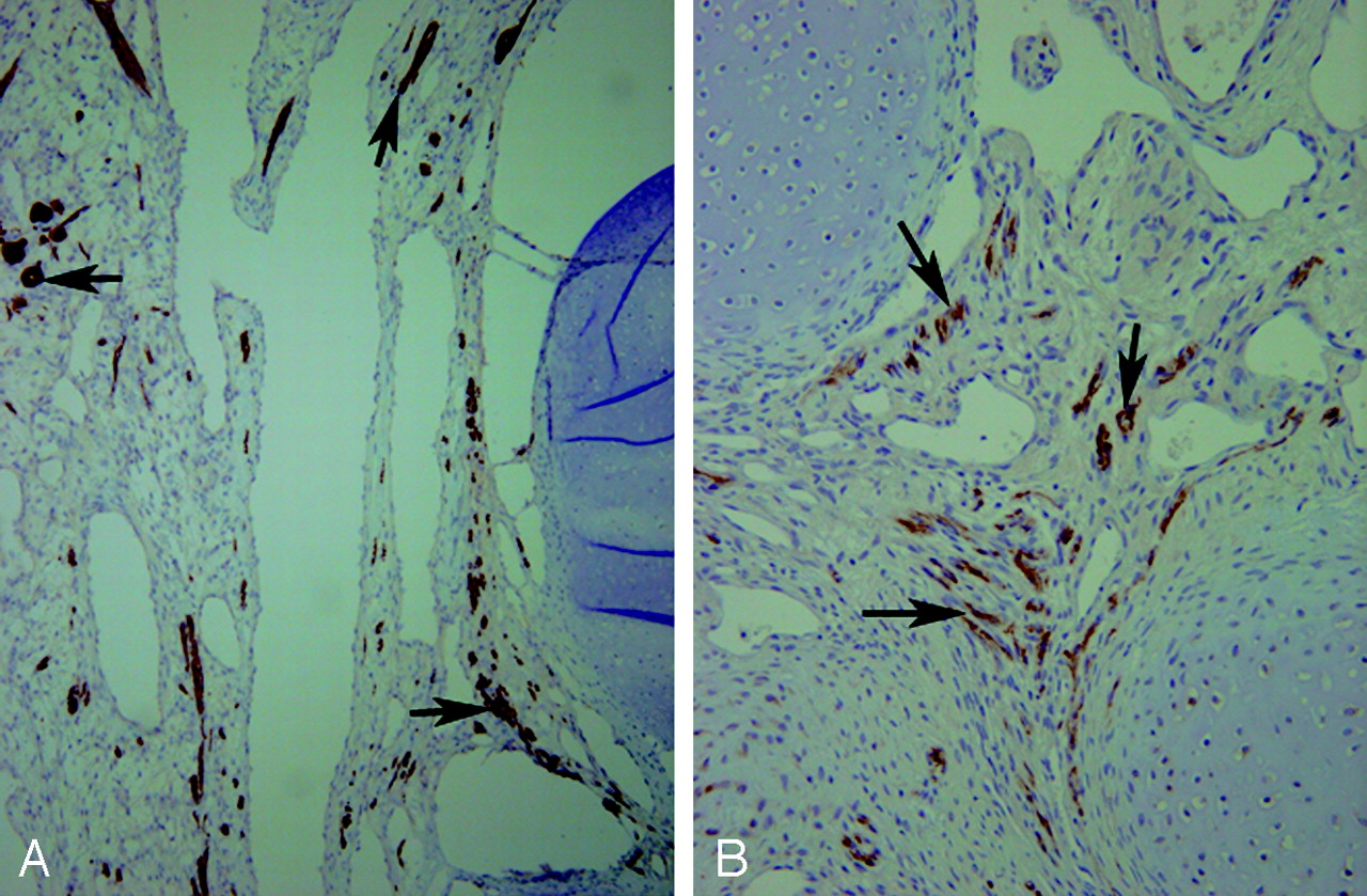
Photomicrographs show immunohistochemical stains for glial fibrillary acidic protein (A) and neurofilament (B) that demonstrate the presence of mature nerve elements (arrows) within the background stroma of the lesion. A, ×40; B, ×200.
Interestingly, a postresection MR imaging, performed 2 days later, demonstrated a residual cervicothoracic intraspinal soft-tissue mass that appeared to be intradural extramedullary in location (Fig 5). Retrospectively, this was also seen on the initial MR imaging. The dura adjacent to this mass appeared to be disrupted, presumably because of the extension of tumor. In reviewing the 2 MR images, we determined that the initial mass had both extradural and intradural-extramedullary components with resection of only the extradural intraspinal component during initial surgery. The patient underwent repeat surgery with reopening of the C4 through T4 laminotomy and complete excision of the residual intradural-extramedullary component. Interestingly, the mass was not attached to the spinal cord and was easily removed in its entirety.
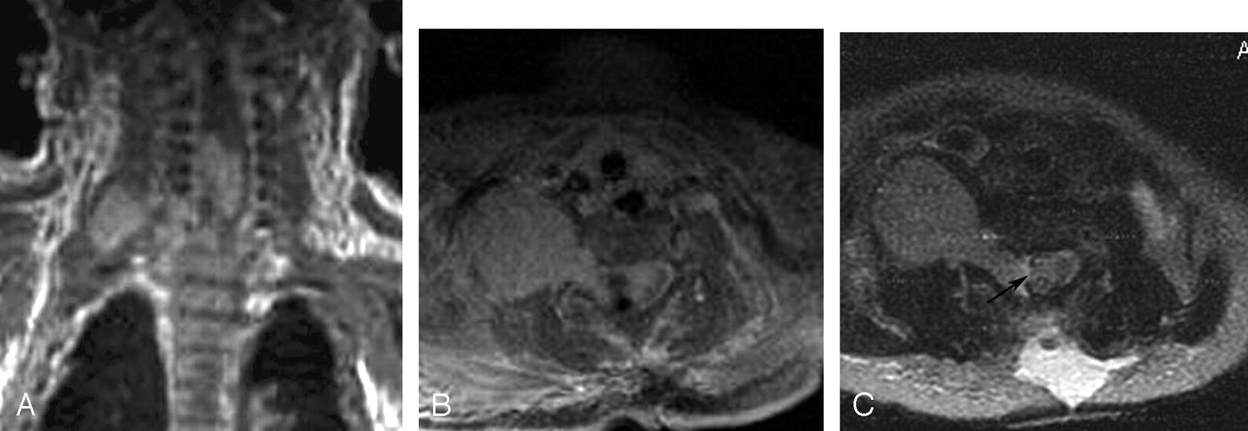
Postoperative (A) axial postcontrast fat-saturated T1-weighted and (B) axial T2-weighted MR images demonstrate a residual well-defined enhancing intradural extramedullary mass. There appears to be a disruption in the dura (arrow).
The child subsequently required several other surgeries, including right diaphragmatic plication for a paralyzed diaphragm, right brachial plexus tumor resection, and right ventriculoperitoneal shunt placement for hydrocephalus. The child was discharged and has been followed clinically and radiologically with no residual or recurrent intraspinal tumor. Some residual weakness in the right upper extremity persists, presumably because of injury to the brachial plexus from the mass effect of the extraspinal component of the tumor and from the surgery.
Discussion
The differential diagnosis for spinal masses in an infant includes spinal dysraphism, congenital tumors, and hamartoma.1 Spinal hamartomas are extremely rare lesions in children, occurring usually intracranially and very rarely in the spine.3 These lesions are composed of mature and well-differentiated elements in an abnormal location.1
Spinal hamartomas are known to present with a skin dimple, cutaneous angioma, subcutaneous mass, or normal overlying skin.1 Our patient presented with a large port wine stain involving the extremity, thorax, and back. According to the existing literature, most patients with spinal hamartomas are neurologically intact at the time of presentation.4 Our patient was a bit unusual because the child presented with upper extremity weakness and decreased movements.
Among congenital spinal tumors, the most common are the teratoma, dermoid, and epidermoid. Teratomas are the most common congenital tumors and contain derivatives of all 3 germ layers by definition (ectoderm, mesoderm, and endoderm), including elements that are heterologous to the site of the lesion.1 These lesions can be benign or malignant and include spinal canal teratomas and sacrococcygeal teratomas.1 Unlike teratomas that contain elements from all the 3 germinal layers, hamartomas are composed of only ectodermal and mesodermal layers (as seen in our patient).4 Dermoids and epidermoids, on the other hand, contain only ectodermal derivatives.4
Neurenteric cyst is an uncommon mass associated with spinal dysraphism. It is a combination of anendodermal (duplication) cyst of foregut origin with a spinal canal dysraphism. Neurenteric cysts are found inthe posterior mediastinum, superior to the carina, and to the right side. These can be differentiated from other masses on the basis of their location and cystic imaging appearance.5
The term “hamartoma” is often used as a generic term to describe any entity with benign characteristics histologically that does not fit into any other general classification. The exact definition in pathologic terms, however, is “an excessive but focal overgrowth of cells and tissues native to the organ in which it occurs. The components of the hamartoma are mature and identical to those found in the remainder of the organ, but are arranged in a disorganized fashion, with aberrant relationships to one another, so that they do not resemble the normal architecture of the tissue from which they arise.”6 Hence, we believed that the use of the term “hamartoma ” was pathologically appropriate in this patient, considering the spinal elements found at tissue biopsy.
Differentiation of spinal masses is essential for the proper surgical approach and management. MR imaging is extremely useful in characterizing these masses and differentiating them. The most interesting feature in our patient was the involvement of 2 compartments of the spinal canal. The initial diagnosis entertained was that of a nerve sheath tumor based on location, foraminal extension, and extraspinal component. However, this patient had a unique finding of an intradural-extramedullary component, which necessitated a second surgery. Had this diagnosis been entertained initially and the intradural component recognized, a second surgery may have been avoided.
Conclusion
The causes of intraspinal masses differ in adults and children. Congenital spinal hamartoma is an extremely rare mass that occurs in children. MR imaging is very useful in demonstrating and characterizing these masses. MR imaging is particularly useful in characterizing the relationship of the mass to the dura, which may result in an alternative diagnosis and, eventually, an altered course of surgical approach and management.
- Received May 18, 2005.
- Accepted after revision June 6, 2005.
- Copyright © American Society of Neuroradiology
In this issue


Jump to section
Related Articles
Cited By...
- No citing articles found.