
Article Figures & Data
Figures
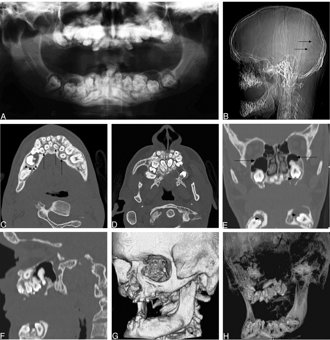
- Fig 1.
A 20-year-old man with pyknodysostosis.
A, Panorex radiograph reveals multiple disorganized crowded deciduous and permanent teeth within the expanded alveolar processes. Many of the teeth are unerupted or only partially erupted. Evaluation of follicles is limited.
B, Scout topogram demonstrates Wormian bones (arrows) within the lambdoid suture. Obtuse gonial angle of the mandible and dental abnormalities are also visualized.
C and D, Axial CT images at the level of the mandible (C) and maxilla (D) reveal multiple unerupted crowded deciduous and permanent teeth within the expanded alveolar processes. Lucencies around several of the teeth have well-defined margins, very likely representing follicles (arrows). However, bandlike zones of demineralization suggest an active inflammatory/infectious process (arrowheads).
E and F, Coronal (E) and sagittal (F) reformatted CT images demonstrate the crowded teeth in the maxilla and mandible with associated lucencies (arrowheads) surrounding the unerupted teeth. Note the hypoplastic maxillary sinuses bilaterally (arrows).
G, Surface-rendered 3D image shows irregularly expanded alveolar processes and mandibular body and a flattened obtuse mandibular angle. The technique limits evaluation for irregular dentition with crowding and retention of deciduous teeth.
H, Volume-rendered 3D image better delineates irregular dentition with crowding and retention of deciduous teeth.


