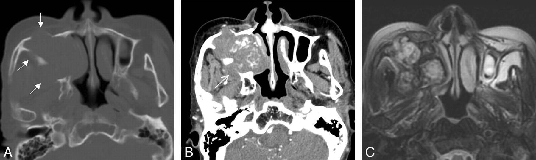
Advertisement

Main menu

  • Home
  • Content
    • Current Issue
    • Publication Preview--Ahead of Print
    • Past Issue Archive
    • Case of the Week Archive
    • Classic Case Archive
    • Case of the Month Archive
  • For Authors
  • About Us
    • About AJNR
    • Editors
    • American Society of Neuroradiology
  • Submit a Manuscript
  • Podcasts
    • Subscribe on iTunes
    • Subscribe on Stitcher
  • More
    • Subscribers
    • Permissions
    • Advertisers
    • Alerts
    • Feedback
  • Other Publications
    • ajnr

User menu

  • Subscribe
  • Alerts
  • Log in

Search

  • Advanced search
American Journal of Neuroradiology

American Journal of Neuroradiology

  • Subscribe
  • Alerts
  • Log in

Advanced Search

  • Home
  • Content
    • Current Issue
    • Publication Preview--Ahead of Print
    • Past Issue Archive
    • Case of the Week Archive
    • Classic Case Archive
    • Case of the Month Archive
  • For Authors
  • About Us
    • About AJNR
    • Editors
    • American Society of Neuroradiology
  • Submit a Manuscript
  • Podcasts
    • Subscribe on iTunes
    • Subscribe on Stitcher
  • More
    • Subscribers
    • Permissions
    • Advertisers
    • Alerts
    • Feedback
  • Follow AJNR on Twitter
  • Visit AJNR on Facebook
  • Follow AJNR on Instagram
  • Join AJNR on LinkedIn
  • RSS Feeds
Research ArticleHEAD & NECK

Sinonasal Organized Hematoma: CT and MR Imaging Findings

E.Y. Kim, H.-J. Kim, S.-K. Chung, H.-J. Dhong, H.Y. Kim, Y.J. Yim, S.T. Kim, P. Jeon and Y.-H. Ko
American Journal of Neuroradiology June 2008, 29 (6) 1204-1208; DOI: https://doi.org/10.3174/ajnr.A1042
E.Y. Kim
  • Find this author on Google Scholar
  • Find this author on PubMed
  • Search for this author on this site
H.-J. Kim
  • Find this author on Google Scholar
  • Find this author on PubMed
  • Search for this author on this site
S.-K. Chung
  • Find this author on Google Scholar
  • Find this author on PubMed
  • Search for this author on this site
H.-J. Dhong
  • Find this author on Google Scholar
  • Find this author on PubMed
  • Search for this author on this site
H.Y. Kim
  • Find this author on Google Scholar
  • Find this author on PubMed
  • Search for this author on this site
Y.J. Yim
  • Find this author on Google Scholar
  • Find this author on PubMed
  • Search for this author on this site
S.T. Kim
  • Find this author on Google Scholar
  • Find this author on PubMed
  • Search for this author on this site
P. Jeon
  • Find this author on Google Scholar
  • Find this author on PubMed
  • Search for this author on this site
Y.-H. Ko
  • Find this author on Google Scholar
  • Find this author on PubMed
  • Search for this author on this site

Fig 2.

Fig 2.
  • Download figure
  • Open in new tab
  • Download powerpoint
Fig 2.

Case 10. Organized hematoma of the maxillary sinus in a 76-year-old woman. A, Precontrast axial CT scan with bone window setting shows a large, expansile soft tissue mass in the right maxillary sinus, which causes cortical thinning and scalloping of bony walls of the maxillary sinus (arrows). With soft tissue windowing, the lesion is isoattenuated to the inferior turbinate with focal hyperattenuated area (data not shown). B, Contrast-enhanced axial CT scan shows marked irregular linear and nodular enhancement within the lesion. Note focal effacement of the retromaxillary fat by the expansile mass that causes smooth erosion of the sinus wall (arrow). C, Axial, fat-suppressed, T2-weighted MR image shows marked heterogeneity of the lesion composed of the areas of hypointensity and isointensity compared with the inferior turbinate. A dark peripheral rim surrounding the lesion is also well demonstrated.

  View article

In this issue

American Journal of Neuroradiology: 42 (1)
American Journal of Neuroradiology
Vol. 42, Issue 1
1 Jan 2021
  • Table of Contents
  • Index by author
  • Complete Issue (PDF)
Print
Download PDF
Email Article

Thank you for your interest in spreading the word on American Journal of Neuroradiology.

NOTE: We only request your email address so that the person you are recommending the page to knows that you wanted them to see it, and that it is not junk mail. We do not capture any email address.

Enter multiple addresses on separate lines or separate them with commas.
Fig 2.
(Your Name) has sent you a message from American Journal of Neuroradiology
(Your Name) thought you would like to see the American Journal of Neuroradiology web site.
CAPTCHA
This question is for testing whether or not you are a human visitor and to prevent automated spam submissions.
Citation Tools
Sinonasal Organized Hematoma: CT and MR Imaging Findings
E.Y. Kim, H.-J. Kim, S.-K. Chung, H.-J. Dhong, H.Y. Kim, Y.J. Yim, S.T. Kim, P. Jeon, Y.-H. Ko
American Journal of Neuroradiology Jun 2008, 29 (6) 1204-1208; DOI: 10.3174/ajnr.A1042

Citation Manager Formats

  • BibTeX
  • Bookends
  • EasyBib
  • EndNote (tagged)
  • EndNote 8 (xml)
  • Medlars
  • Mendeley
  • Papers
  • RefWorks Tagged
  • Ref Manager
  • RIS
  • Zotero
Share
Sinonasal Organized Hematoma: CT and MR Imaging Findings
E.Y. Kim, H.-J. Kim, S.-K. Chung, H.-J. Dhong, H.Y. Kim, Y.J. Yim, S.T. Kim, P. Jeon, Y.-H. Ko
American Journal of Neuroradiology Jun 2008, 29 (6) 1204-1208; DOI: 10.3174/ajnr.A1042
del.icio.us logo Digg logo Reddit logo Twitter logo CiteULike logo Facebook logo Google logo Mendeley logo
  • Tweet Widget
  • Facebook Like
  • Google Plus One

Related Articles

  • PubMed
  • Google Scholar

Cited By...

  • MR Evaluation of Sinonasal Angiomatous Polyp
  • Apparent Diffusion Coefficient Mapping for Sinonasal Diseases: Differentiation of Benign and Malignant Lesions
  • Crossref
  • Google Scholar

This article has not yet been cited by articles in journals that are participating in Crossref Cited-by Linking.

More in this TOC Section

  • Skull Base Osteomyelitis: A Comprehensive Imaging Review
  • The Many Faces of Persistent Stapedial Artery: CT Findings and Embryologic Explanations
  • Imaging Parameters of the Ipsilateral Medial Geniculate Body May Predict Prognosis of Patients with Idiopathic Unilateral Sudden Sensorineural Hearing Loss on the Basis of Diffusion Spectrum Imaging
Show more HEAD & NECK

Similar Articles

Advertisement

News and Updates

  • Lucien Levy Best Research Article Award
  • Thanks to our 2020 Distinguished Reviewers
  • Press Releases

Resources

  • Evidence-Based Medicine Level Guide
  • How to Participate in a Tweet Chat
  • AJNR Podcast Archive
  • Ideas for Publicizing Your Research
  • Librarian Resources
  • Terms and Conditions

Opportunities

  • Share Your Art in Perspectives
  • Get Peer Review Credit from Publons
  • Moderate a Tweet Chat

American Society of Neuroradiology

  • Neurographics
  • ASNR Annual Meeting
  • Fellowship Portal
  • Position Statements

© 2021 by the American Society of Neuroradiology | Print ISSN: 0195-6108 Online ISSN: 1936-959X

Powered by HighWire