
Article Figures & Data
Figures
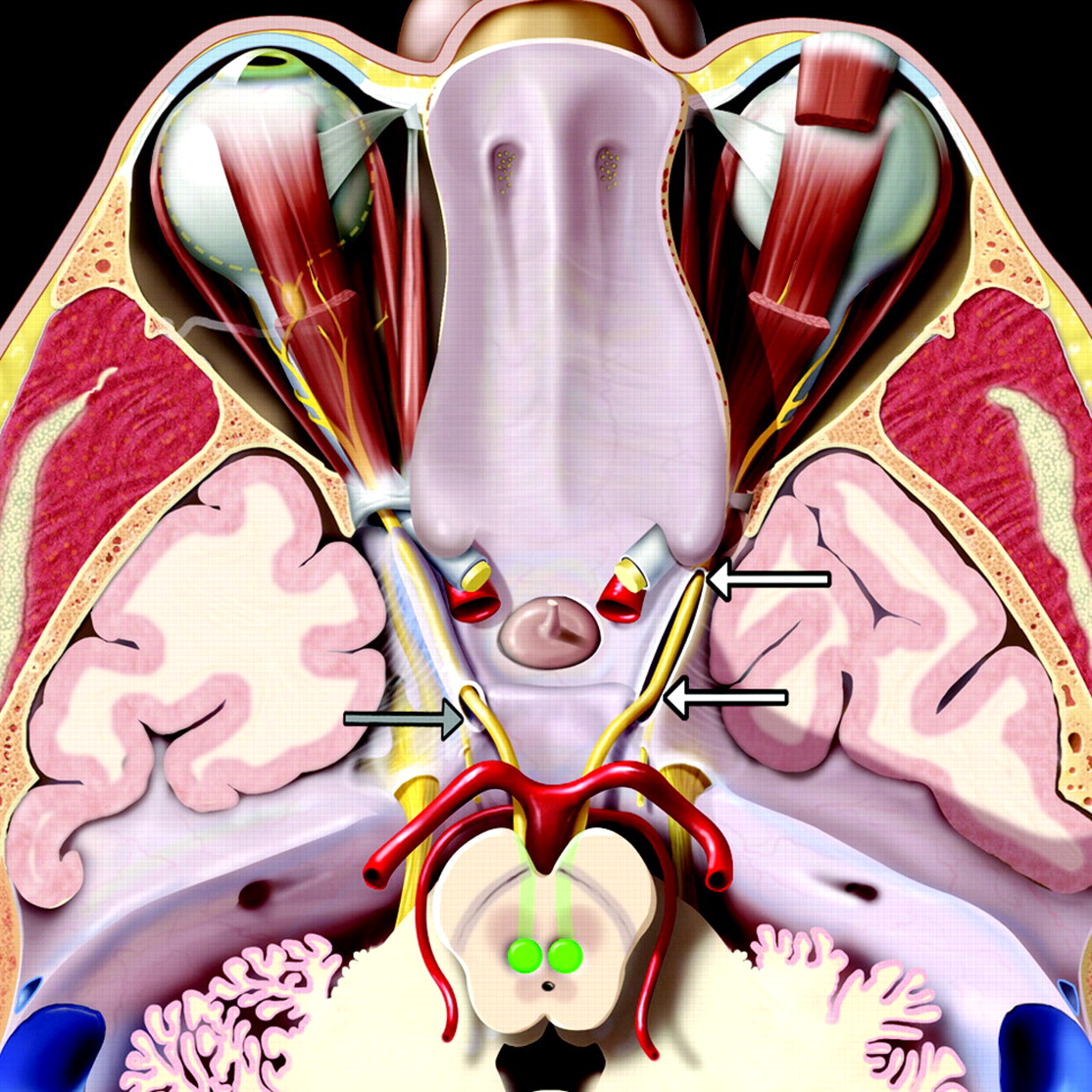
- Fig 1.
An axial graphic of the oculomotor nerves (CIIIs), seen from above, shows their nuclei (green) in front of the periaqueductal gray matter. Nerve fibers course anteriorly across the red nuclei and substantia nigra and exit the midbrain at the interpeduncular fossa. They then run anterolaterally between the posterior cerebral and superior cerebellar arteries. The nerves enter the cavernous sinus roof at the obliquely oriented porus (gray arrow). Each nerve is accompanied by an arachnoid-lined CSF-filled sleeve (the OMC). The OMC surrounding the right CNIII (white arrows) has been opened to depict the nerve as it courses anteriorly within the cistern. The OMCs terminate near the anterior clinoid process as the CNIIIs enter the orbit through the superior orbital fissures.
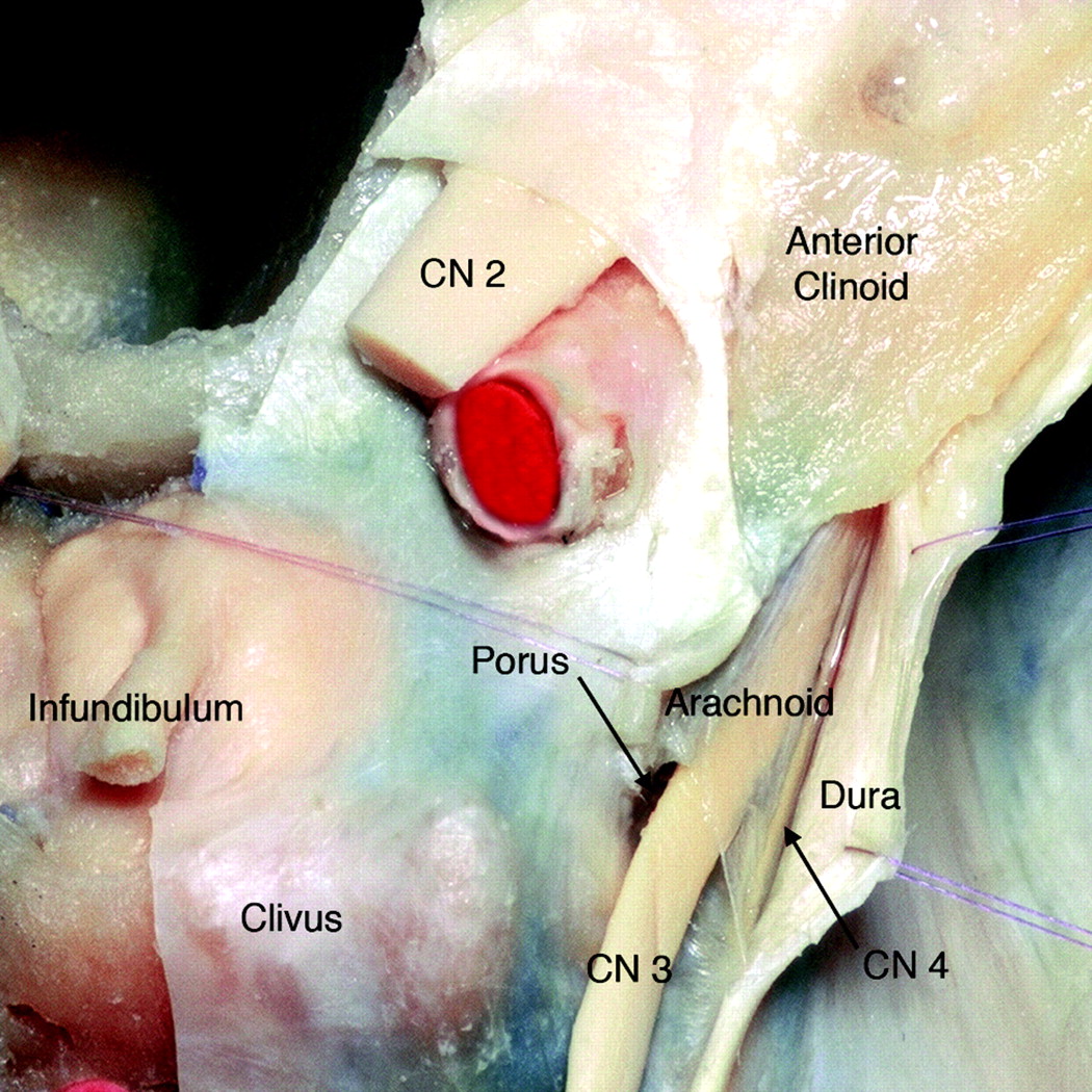
- Fig 2.
Axial anatomic dissection, seen from above, shows the relationship of the right CNIII to the clivus and cavernous sinus dura. The dura is dissected and retracted laterally to expose the arachnoid-lined CSF-filled OMC as it runs toward the anterior clinoid. Note the oblique orientation of the porus (modified and reprinted with permission from Martins et al1).
- Fig 3.
Sagittal anatomic dissection of the cavernous sinus with the dura partially removed and the oculomotor nerve exposed. The lateral wall of the OMC has been removed to depict the nerve within the cistern. The OMC is outlined in green. The CNIII does not become incorporated into the fibrous dural cavernous sinus wall until it reaches the lower margin of the anterior clinoid process. Meckel cave is outlined in pink (modified and reprinted with permission from Martins et al1).
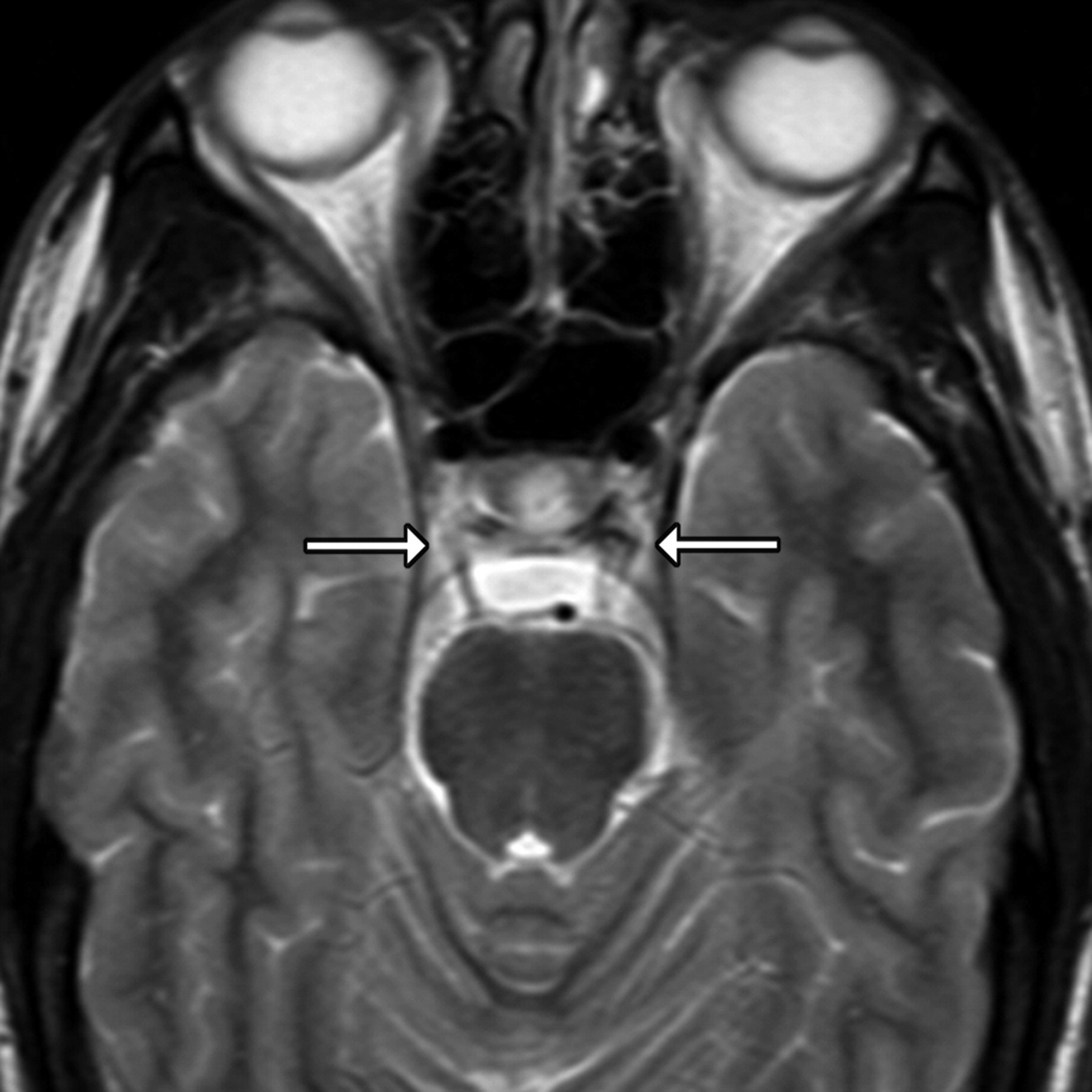
- Fig 4.
Axial T2-weighted image of a routine brain scan shows bilateral OMCs (arrows).
- Fig 5.
A, Axial T2-weighted image from a routine high-resolution 3T screening study to evaluate internal auditory canal lesions shows the right CNIII entering the porus and the left CNIII within the CSF-filled OMC (arrows). B, Coronal T2-weighted image from a routine high-resolution 3T screening study to evaluate IAC lesions shows CNIIIs in the CSF-filled OMCs (arrows).
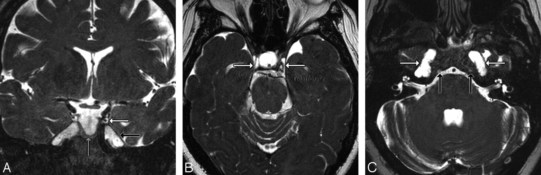
- Fig 6.
A, Dedicated volunteer axial 3T CISS scan shows CNIIIs and OMCs. Note that each nerve (black arrow) enters its OMC through the obliquely oriented porus, seen here as horseshoe-shaped CSF collections (gray arrow) just lateral to the clivus and posterior clinoid processes. Each OMC (white arrow) continues anterolaterally toward the anterior clinoid process. B, Dedicated volunteer coronal 3T CISS scan through the middle of the cavernous sinus shows the CNIIIs within the OMCs. The appearance of isointense “dots” surrounded by hyperintense “rings” of CSF is the most easily identified appearance of the OMC and CNIII (arrows). C, Dedicated volunteer sagittal 3T CISS reformatted from the axial data shows the entire course of CNIII (black arrow) from its exit at the interpeduncular fossa, through the prepontine cistern, and its entrance into the OMC (white arrow) at the porus (gray arrow).
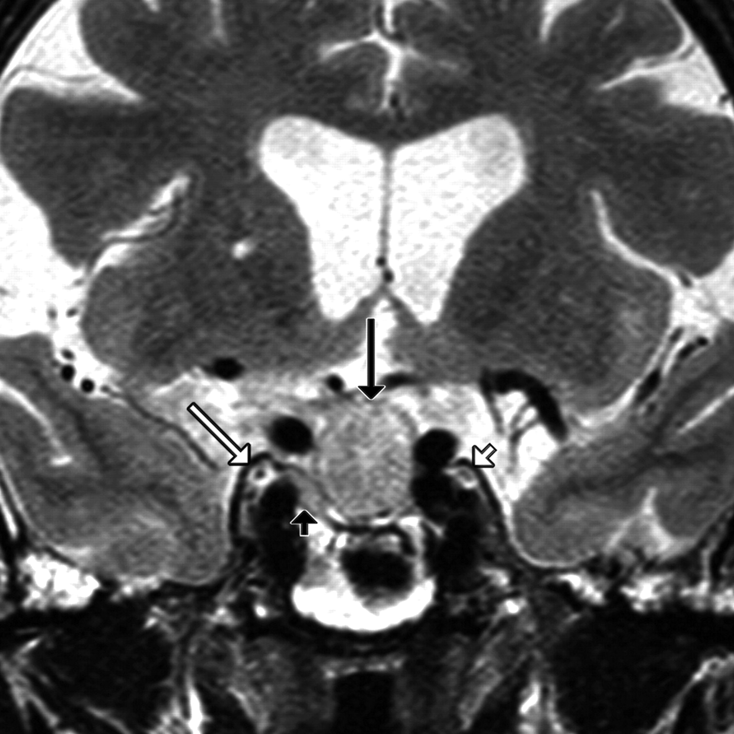
- Fig 7.
Coronal T2-weighted image in a patient with a pituitary macroadenoma (black arrow) shows superolateral extension of the tumor (black arrowhead) toward the CSF-filled OMC (white arrow). Note that the tumor abuts the OMC but does not invade it. The left OMC and CNIII are normal (white arrowhead).
- Fig 8.
Axial T2-weighted image in a patient with diplopia shows the normal CSF-filled OMC surrounding the left third oculomotor nerve (white arrow). An isointense clival meningioma (black arrow) encroaches on the lateral aspect of the right OMC (white arrowhead), effacing the CSF but sparing the third nerve.
- Fig 9.
A, Coronal T2-weighted image in a patient with generalized dural ectasia shows enlarged OMCs (white arrow), a partial empty sella (black arrow), and patulous Meckel caves (gray arrow). B, Axial T2-weighted image in the same patient shows strikingly enlarged OMCs (arrows). C, A lower section through the IACs shows bilaterally enlarged Meckel caves (white arrows) and Dorello canals (black arrows), part of the generalized dural ectasia. The IACs appear normal.


