
Article Figures & Data
Figures
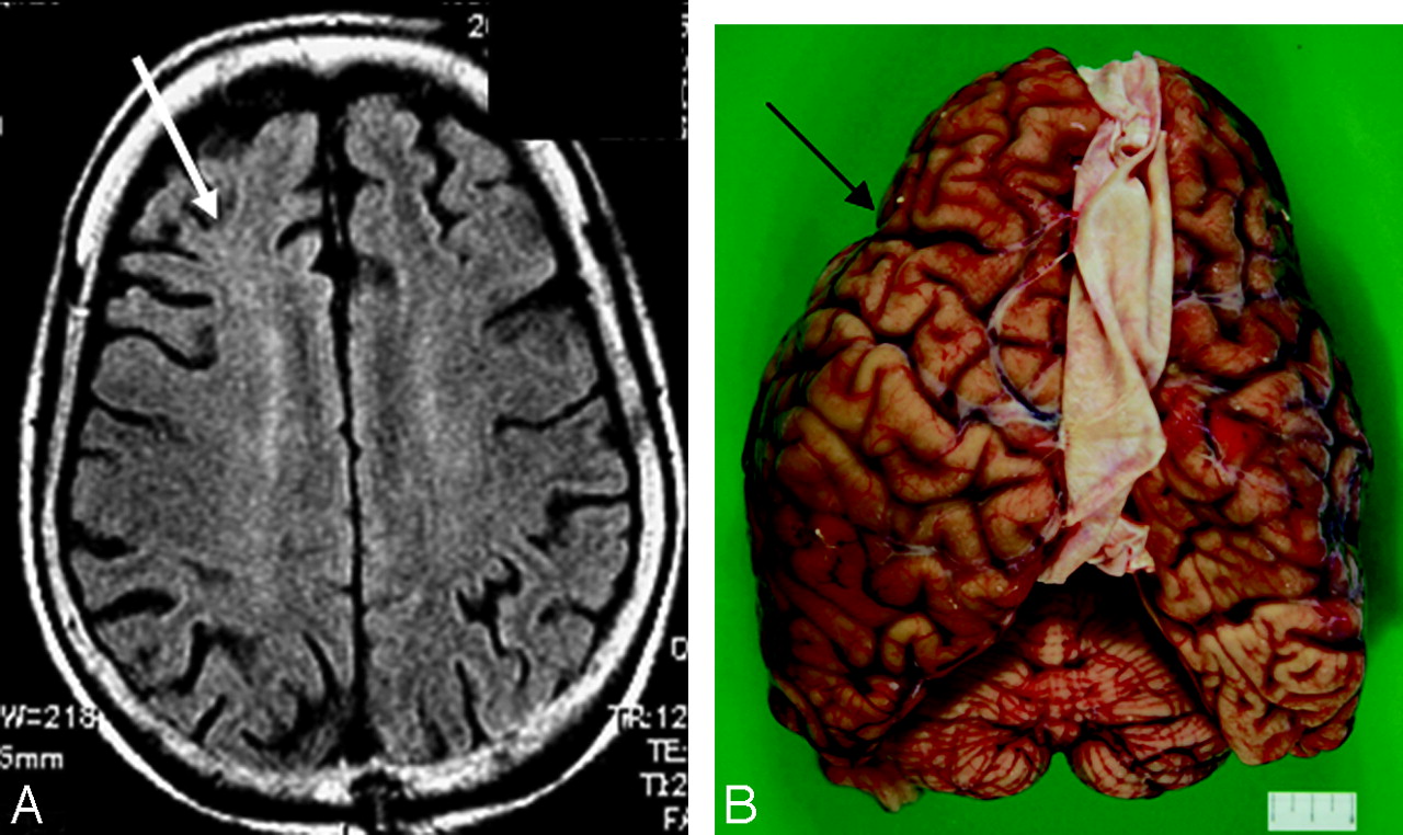
- Fig 1.
Corticobasal degeneration, case 1. An 84-year-old woman. A, Axial T2-weighted image shows right-side-dominant atrophy including the central sulcus (arrow). B, A macrospecimen of this patient shows right-frontal-dominant atrophy (arrow).
- Fig 2.
Corticobasal degeneration, case 2. A 74-year-old woman. A, An axial fluid-attenuated inversion recovery image 3 years before autopsy shows no obvious asymmetric atrophy. Subcortical hyperintensity is shown in the right frontal white matter (white arrow). B, A macrospecimen of this patient shows mild frontal atrophy with some asymmetry (arrow).
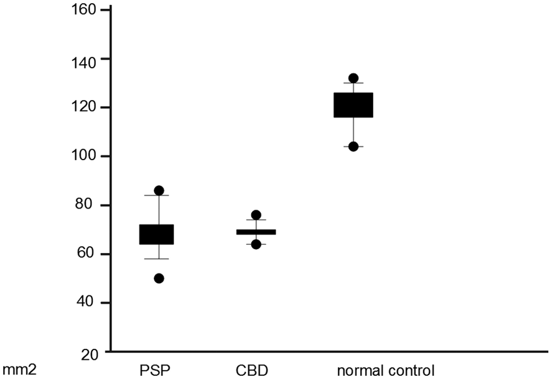
- Fig 3.
Scatterplot (mean, SD, and range) of the area of the midbrain in patients with progressive supranuclear palsy (PSP), corticobasal degeneration (CBD), and age-matched healthy controls. There was no individual overlap of the midbrain tegmental area between the healthy controls and patients with CBD and PSP, apparently showing that severe atrophy of the midbrain tegmentum was present in patients with CBD and PSP.
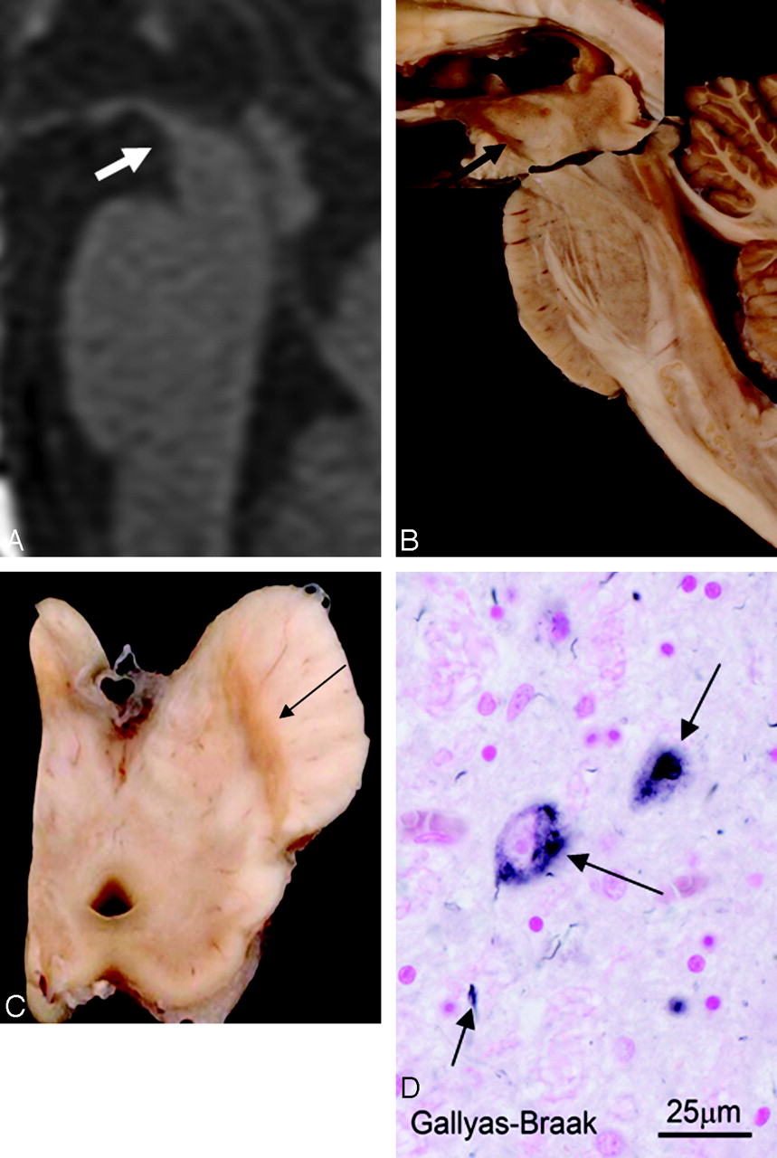
- Fig 4.
Corticobasal degeneration (CBD), case 1. An 84-year-old woman. A, T1-weighted midsagittal image clearly shows atrophy of the midbrain tegmentum (arrow). The area of the midbrain tegmentum is 73 mm2. B, A macroscopic specimen of the midbrain shows marked atrophy (arrow). C, A macroscopic view of the midbrain shows discoloration of the substantia nigra (arrow). D, A microscopic view of the substantia nigra (Gallyas-Braak stain) shows argyrophilic threads and granular or fibrous inclusion bodies (arrows). These are consistent with CBD.
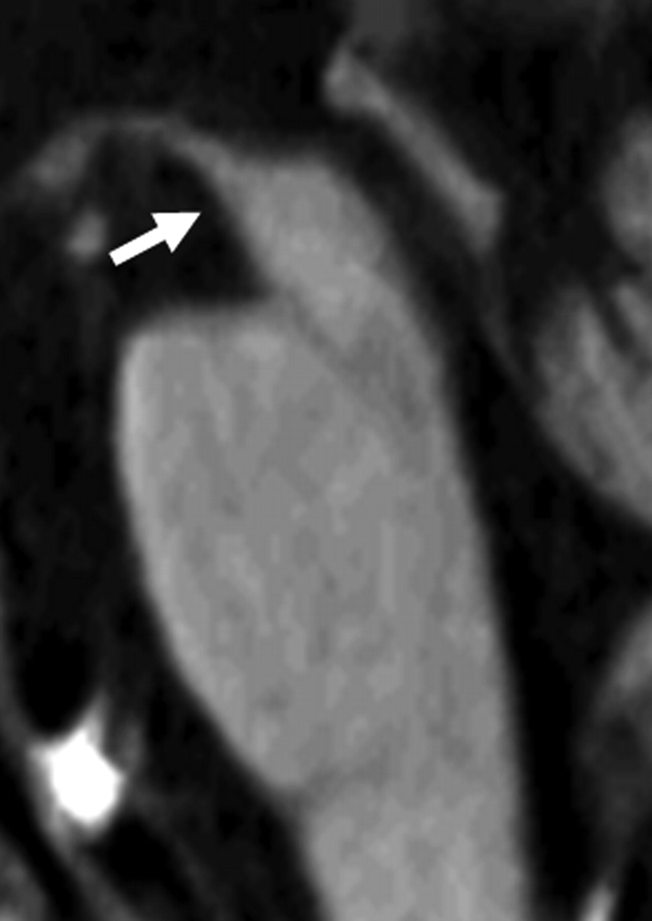
- Fig 5.
An age-matched healthy control 72-year-old woman. T1-weighted midsagittal image shows no obvious atrophy of the midbrain tegmentum (arrow). The area of the midbrain tegmentum is 128 mm2.
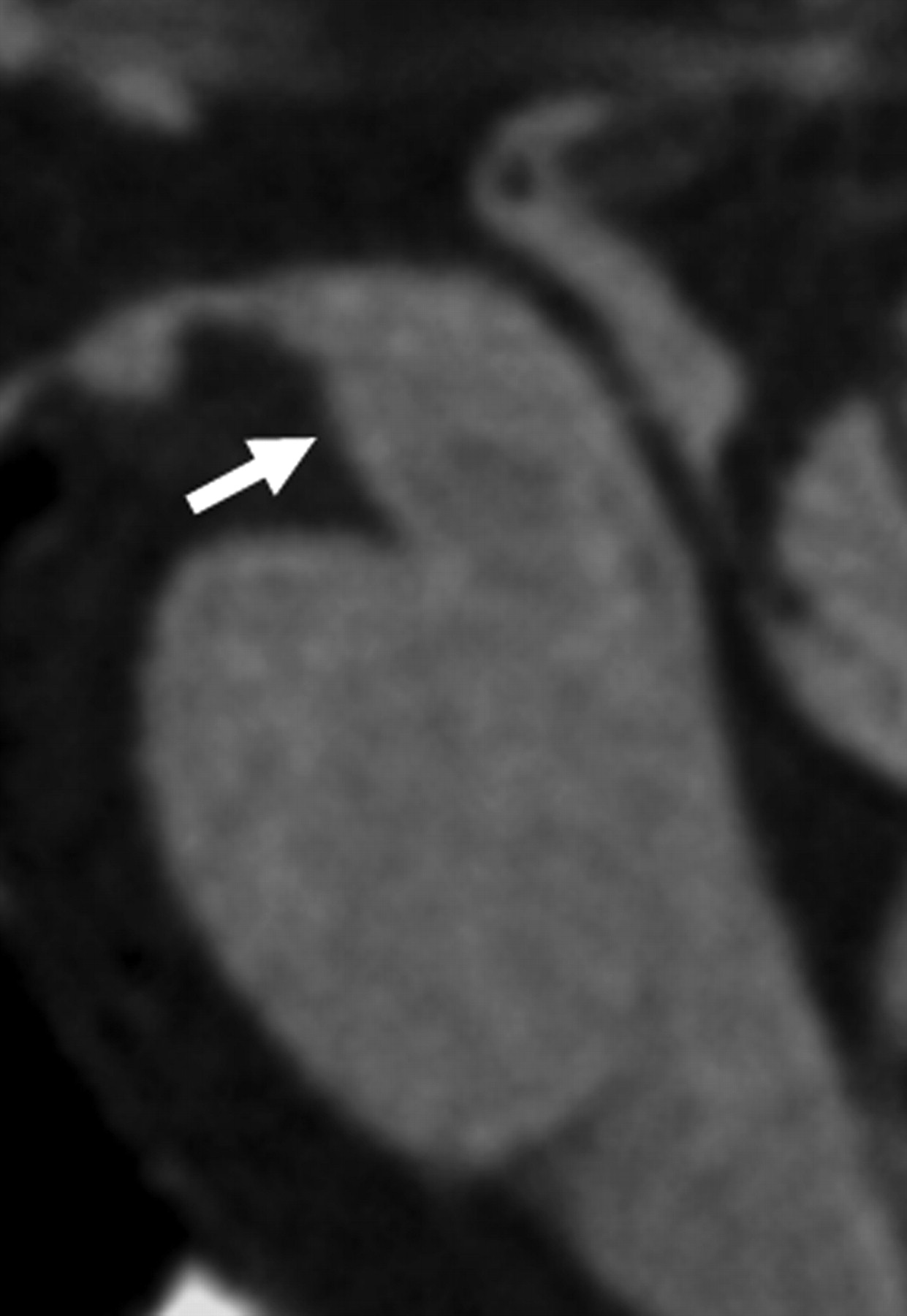
- Fig 6.
Progressive supranuclear palsy. A 74-year-old man. T1-weighted midsagittal image clearly shows atrophy of the midbrain tegmentum (arrow). The area of the midbrain tegmentum is 71 mm2.
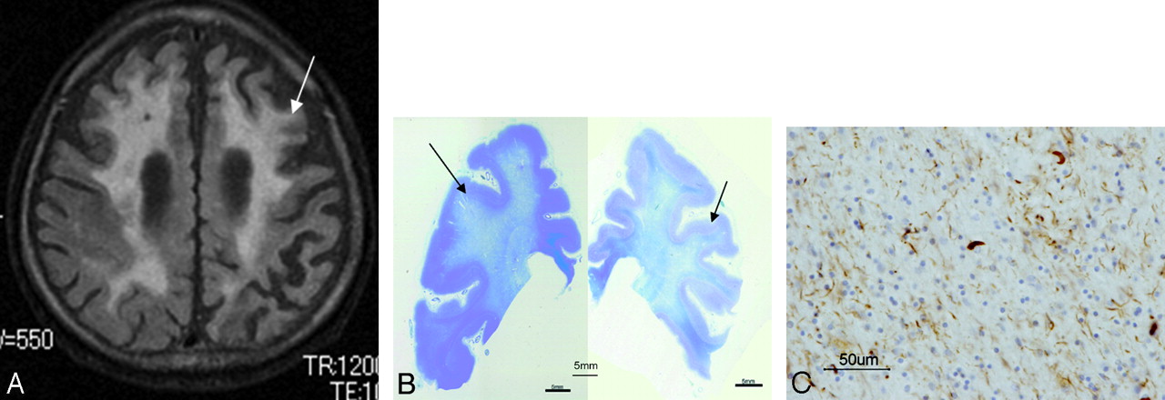
- Fig 7.
Corticobasal degeneration (CBD), case 1. An 84-year-old woman. A, Axial T2-weighted image shows a high signal intensity in the right frontal subcortical white matter (white arrow). B, In a microscopic specimen of the right frontal lobe corresponding to the site of white matter lesions, myelin sheath staining is decreased (red oval). The scale is 1 mm. C, In this area, there is positive staining for antiphosphorylated tau antibody on AT8 staining, which is compatible with the primary changes in CBD. The scale is 50 μm.
- Fig 8.
Corticobasal degeneration (CBD), case 3. A 70-year-old man. A, An axial fluid-attenuated inversion recovery image shows a high signal intensity bilaterally over a wide area in the frontal lobes (arrow). B, Corresponding to sites of white matter lesions, myelin sheath staining is decreased (arrow). The scale is 5 mm. C, These sites are stained positively for antiphosphorylated tau antibody. The scale is 50 μm. The changes are primary characteristics of CBD.
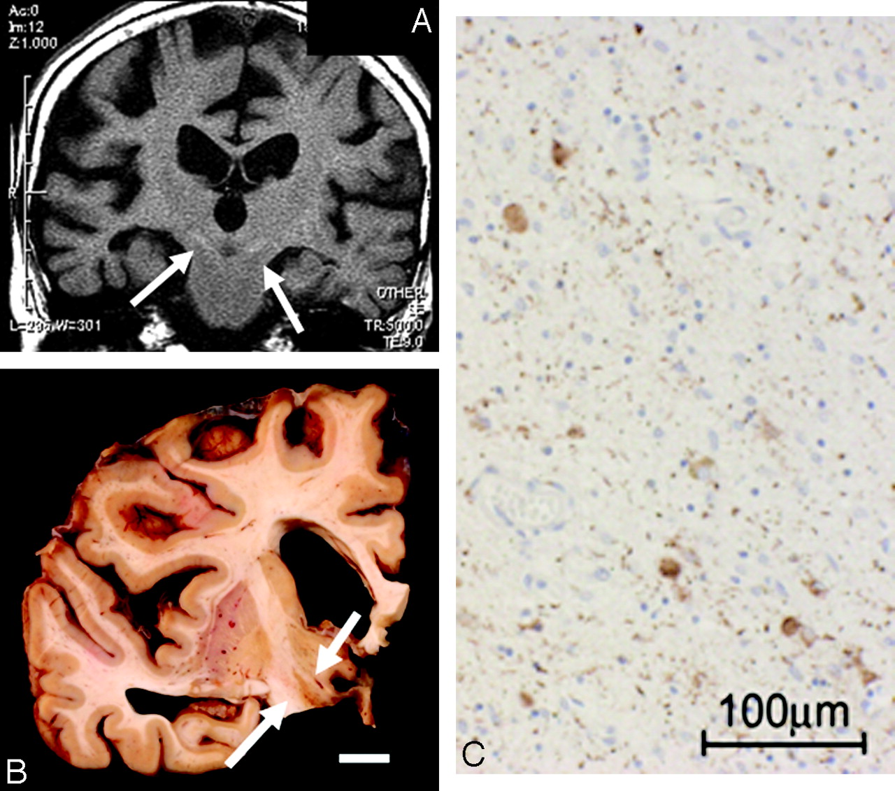
- Fig 9.
Corticobasal degeneration (CBD), case 1. An 84-year-old woman. A, Coronal T1-weighted image shows symmetric high signal intensity bilaterally in the subthalamic nuclei (arrows).B, A macroscopic specimen shows a brownish change in the subthalamic nuclei (arrows). C, On microscopic examination (AT8 stain), antiphosphorylated tau antibody–positive neurons and gliosis are observed. These changes are characteristic of CBD. The scale is 100 μm.
- Fig 10.
Progressive supranuclear palsy (PSP). An 84-year-old man. A, Coronal T1-weighted image shows a symmetric high signal intensity bilaterally in the subthalamic nuclei (arrow). B, A microscopic specimen of myelin-sheath staining shows the atrophic change of the subthalamic nuclei (arrows). The scale is 1 mm. C, On microscopic examination in the subthalamic nuclei, AT8 staining is clearly positive in the neurons (brown area). An enlarged image shows a tuft. These changes are characteristic of PSP. The scale is 100 μm.


Tables
Case No. Age at Onset (yr) Sex Duration (yr) Rigidity Dystonia Pyramidal Signs Cortical Dysfunction Vertical Gaze Palsy Dementia CDx 1 74 F 10 Lt>Rt – – Ocular apraxia + Mute PSP? 2 68 F 6 Lt>Rt Apraxia (Lt hand) ± + Severe akinetic mute AD 3 67 M 3 Rt>Lt No cortical sign + + Severe PDD 4 74 F 6 Rt>Lt – – – + + Severe CBD Note:—CBD indicates corticobasal degeneration; PSP?, progressive supranuclear palsy suspected; CDx, clinical diagnosis; AD, Alzheimer disease; PDD, Parkinson disease with dementia; –, no symptom; +, obvious symptom; ±, suspicious symptom; Lt, left; Rt, right.
Case No. Atrophy (Dominant Cerebral Hemisphere) White Matter Hyperintensity on FLAIR Hyperintensity on T1WI in Bil Subthalamic Nucleus Precentral Gyrus Frontal Lobe 1 Rt frontal operculum and convexity – + + 2 Bil frontal convexity – + + 3 Lt frontoparietal Bil + + 4 Rt frontoparietal Rt + + Note:—FLAIR indicates fluid-attenuated inversion recovery; T1WI = T1-weighted imaging; Bil, bilateral; –, no signal abnormality; +, obvious signal abnormality.


