Advertisement

Main menu

  • Home
  • Content
    • Current Issue
    • Publication Preview--Ahead of Print
    • Past Issue Archive
    • Case of the Week Archive
    • Classic Case Archive
    • Case of the Month Archive
    • COVID-19 Content and Resources
  • For Authors
  • About Us
    • About AJNR
    • Editors
    • American Society of Neuroradiology
  • Submit a Manuscript
  • Podcasts
    • Subscribe on iTunes
    • Subscribe on Stitcher
  • More
    • Subscribers
    • Permissions
    • Advertisers
    • Alerts
    • Feedback
  • Other Publications
    • ajnr

User menu

  • Subscribe
  • Alerts
  • Log in

Search

  • Advanced search
American Journal of Neuroradiology

American Journal of Neuroradiology

  • Subscribe
  • Alerts
  • Log in

Advanced Search

  • Home
  • Content
    • Current Issue
    • Publication Preview--Ahead of Print
    • Past Issue Archive
    • Case of the Week Archive
    • Classic Case Archive
    • Case of the Month Archive
    • COVID-19 Content and Resources
  • For Authors
  • About Us
    • About AJNR
    • Editors
    • American Society of Neuroradiology
  • Submit a Manuscript
  • Podcasts
    • Subscribe on iTunes
    • Subscribe on Stitcher
  • More
    • Subscribers
    • Permissions
    • Advertisers
    • Alerts
    • Feedback
  • Follow AJNR on Twitter
  • Visit AJNR on Facebook
  • Follow AJNR on Instagram
  • Join AJNR on LinkedIn
  • RSS Feeds
Research ArticleBRAIN

Territorial and Microvascular Perfusion Impairment in Brain Arteriovenous Malformations

J. Fiehler, T. Illies, M. Piening, D. Säring, N. Forkert, J. Regelsberger, U. Grzyska, H. Handels and J.V. Byrne
American Journal of Neuroradiology February 2009, 30 (2) 356-361; DOI: https://doi.org/10.3174/ajnr.A1351
J. Fiehler
  • Find this author on Google Scholar
  • Find this author on PubMed
  • Search for this author on this site
T. Illies
  • Find this author on Google Scholar
  • Find this author on PubMed
  • Search for this author on this site
M. Piening
  • Find this author on Google Scholar
  • Find this author on PubMed
  • Search for this author on this site
D. Säring
  • Find this author on Google Scholar
  • Find this author on PubMed
  • Search for this author on this site
N. Forkert
  • Find this author on Google Scholar
  • Find this author on PubMed
  • Search for this author on this site
J. Regelsberger
  • Find this author on Google Scholar
  • Find this author on PubMed
  • Search for this author on this site
U. Grzyska
  • Find this author on Google Scholar
  • Find this author on PubMed
  • Search for this author on this site
H. Handels
  • Find this author on Google Scholar
  • Find this author on PubMed
  • Search for this author on this site
J.V. Byrne
  • Find this author on Google Scholar
  • Find this author on PubMed
  • Search for this author on this site
  • Article
  • Figures & Data
  • Info & Metrics
  • References
  • PDF
Loading

Article Figures & Data

Figures

  • Tables
  • Fig 1.
    • Download figure
    • Open in new tab
    • Download powerpoint
    Fig 1.

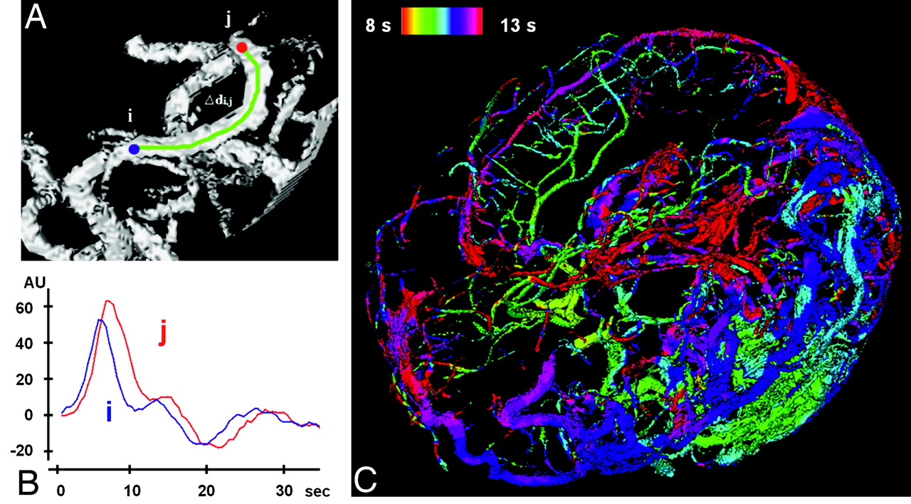
    Temporal assignment of inflow time points. A, Section of an MIP of a vessel tree (based on 4D-MRA) with 2 manually selected locations (i and j). B, In maximum slope (in AUs) versus time (in seconds) curves, the time points for the maximum slope can be determined separately for i and j, and a delay can be calculated. C, The time points of those maxima are color-coded and projected onto the complete vessel tree of the AVM in the TOF-MRA.

  • Fig 2.
    • Download figure
    • Open in new tab
    • Download powerpoint
    Fig 2.

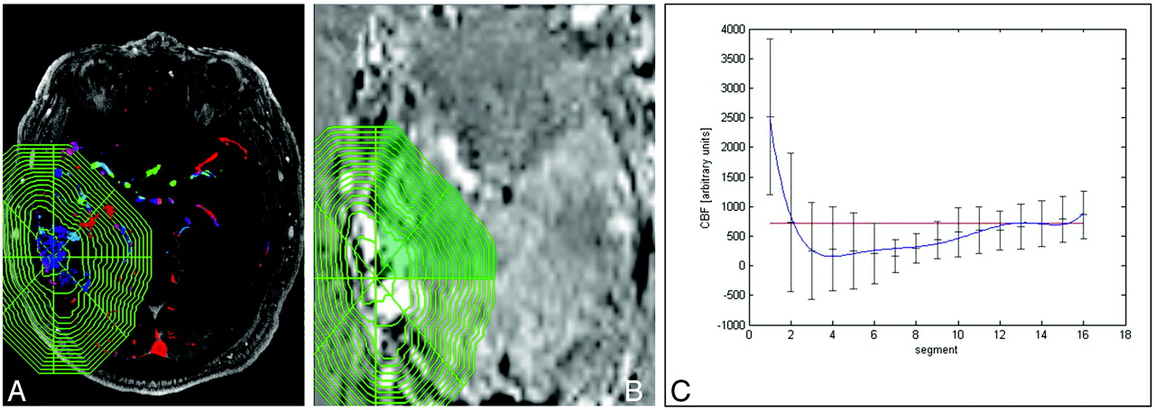
    A and B, Determination of the PND after manual delineation of the AVM nidus in the TOF-MRA source images (A) and after the complex shape is transferred to the coregistered PASL map (B). C, Signal intensities for each piece of cake are determined in AUs in each segment beginning from the AVM nidus to the periphery. The segments outside the skull are excluded from further analysis. s indicates seconds.

  • Fig 3.
    • Download figure
    • Open in new tab
    • Download powerpoint
    Fig 3.

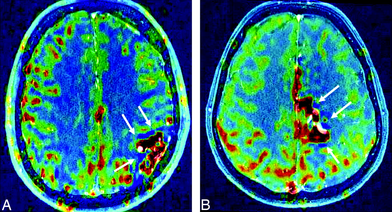
    PASL maps (CBF-weighted) are coregistered to TOF-MRA source images. The patients either fulfill the 20% dip criterion (A, patient 18; 3647) or do not (B, patient 13; 3136). Nevertheless, dark-blue low CBF regions (arrows) are observed in both patients, suggesting local CBF decreases, possibly as a result of a local steal. Red indicates high CBF; dark blue, low CBF.
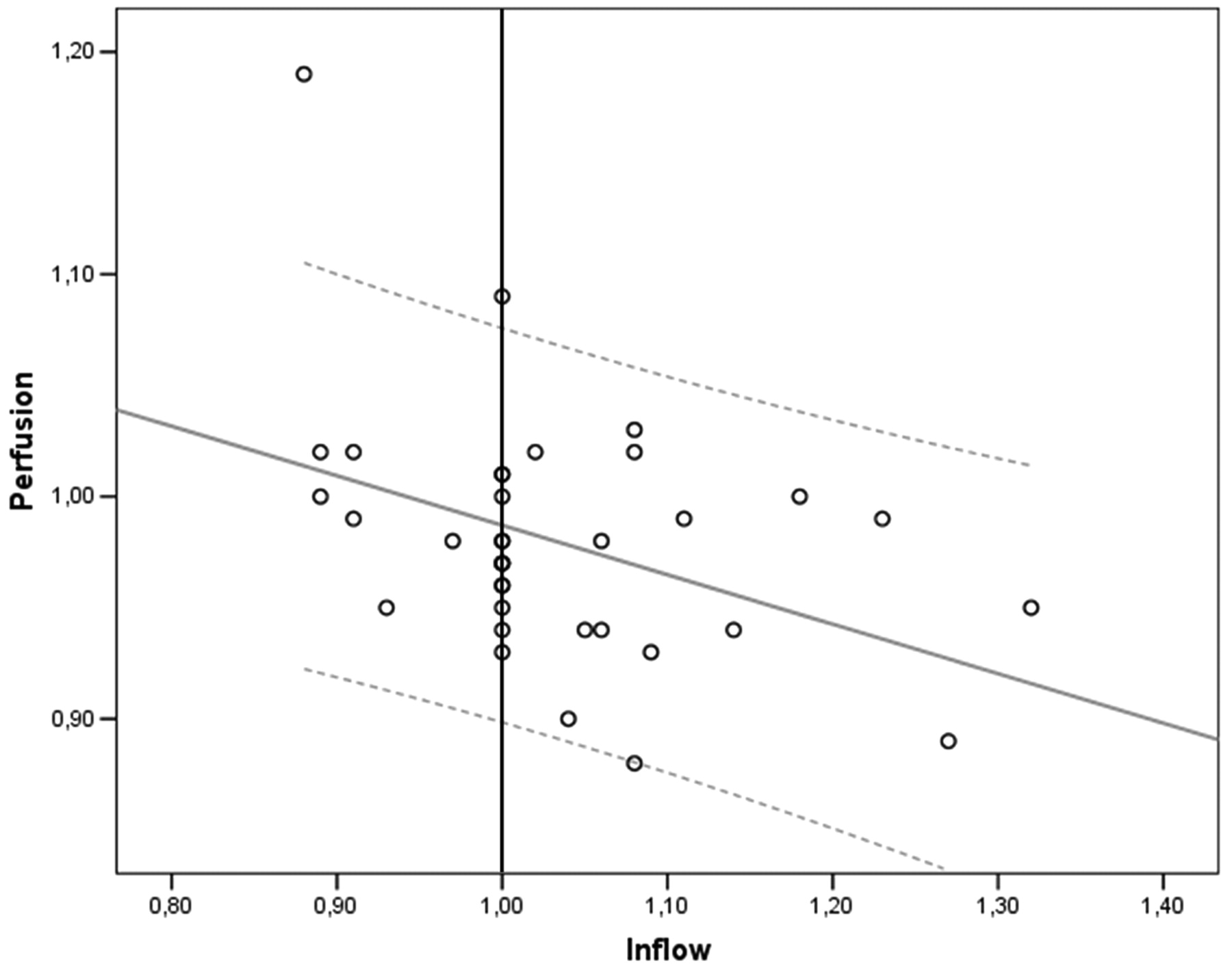
  • Fig 4.
    • Download figure
    • Open in new tab
    • Download powerpoint
    Fig 4.

    There is a negative correlation of the perfusion ratio with the inflow ratio in a combined analysis of all affected vascular territories (R = −0.402, P = .015). The gray line represents the regression line with 95% confidence intervals (dotted lines).

Tables

  • Figures
    • View popup
    Table 1:

    Demographics

    No.AgeSexNidal Volume (mL)Feeding ArterySpetzler Grade20CorticalLocationSideSymptoms
    ACAMCAPCA
    155M40–++4YesTemporalRSAH >6 months ago
    233M300+++4YesParieto-occipitalLSeizures + headache
    342M330–++5YesTemporo-occipitalRSAH >6 months ago
    451M130++–3YesFrontalLTinnitus left side pronounced
    545M130––+4YesOccipitalLSeizures + headache
    628M50+++5YesTemporalLSeizures + stuttering
    733F270+++4YesFrontoparietalRSeizures
    822F20–++3YesOccipitotemporalRSeizures + visual field defects
    922M70–+–3NoTemporalRSeizures
    1017F100+––3NoFrontoparietalLIncidental
    1135F340++–2YesParietalLSeizures + dysaesthesia
    1247M80–++3YesTemporalLSeizures
    1341F20++–2YesParietalLPulsatile headache
    1451F40–+–3NoBasal gangliaLAphasia
    1545M40+–+3YesFrontoparietalL+RIntraventricular hemorrhage >6 months ago
    1652M10––+2YesParapontineRSAH >6 months ago
    1731F30–+–3YesFrontalRSeizures
    1840M10–++4YesTemporalLSeizures
    1922M120–++1YesTemporoparietalLSeizures
    2019M60+–+5NoCorpus callosumLIncidental
    • Note:—SAH indicates subarachnoid hemorrhage; R, right, L, left; –, not involved; +, involved; ACA, anterior cerebral artery; MCA, middle cerebral artery; PCA, posterior cerebral artery.

    • View popup
    Table 2:

    Perfusion ratios of affected-versus-nonaffected hemispheres for each vascular territory

    Patient No.ACAMCAPCAPND (%)Visible
    11.010.951.02–Yes
    21.020.980.99–Yes
    31.020.940.88–Yes
    4–1.001.09–Yes
    50.930.960.90–Yes
    60.980.931.00–Yes
    70.960.950.90–No
    81.031.000.89–Yes
    90.990.940.94–Yes
    100.960.950.9049Yes
    111.001.010.89–Yes
    121.190.980.9439No
    130.960.970.93–Yes
    140.950.980.9752Yes
    150.990.990.99–No
    161.091.021.02–Yes
    171.011.000.99–Yes
    180.990.981.0262Yes
    190.970.931.01–Yes
    200.980.920.8278Yes
    • Note:—PND indicates perinidal dip.

    • View popup
    Table 3:

    Inflow times at predefined measuring points

    Affected Hemisphere (in seconds)Contralateral Hemisphere (in seconds)Ratio Affected/ Contralateral
    ICA10.8 ± 3.911.9 ± 4.20.96 ± 0.19
    MCA9.2 ± 2.59.5 ± 2.81.00 ± 0.33
    ACA10.2 ± 2.510.3 ± 2.70.94 ± 0.23
    PCA12.0 ± 5.012.4 ± 4.90.90 ± 0.16
    TS11.8 ± 3.011.6 ± 3.61.09 ± 0.94
    • Note:—ICA indicates internal carotid artery; TS, transverse sinus.

PreviousNext
Back to top

In this issue

American Journal of Neuroradiology: 30 (2)
American Journal of Neuroradiology
Vol. 30, Issue 2
February 2009
  • Table of Contents
  • Index by author
Advertisement
Print
Download PDF
Email Article

Thank you for your interest in spreading the word on American Journal of Neuroradiology.

NOTE: We only request your email address so that the person you are recommending the page to knows that you wanted them to see it, and that it is not junk mail. We do not capture any email address.

Enter multiple addresses on separate lines or separate them with commas.
Territorial and Microvascular Perfusion Impairment in Brain Arteriovenous Malformations
(Your Name) has sent you a message from American Journal of Neuroradiology
(Your Name) thought you would like to see the American Journal of Neuroradiology web site.
CAPTCHA
This question is for testing whether or not you are a human visitor and to prevent automated spam submissions.
Citation Tools
Territorial and Microvascular Perfusion Impairment in Brain Arteriovenous Malformations
J. Fiehler, T. Illies, M. Piening, D. Säring, N. Forkert, J. Regelsberger, U. Grzyska, H. Handels, J.V. Byrne
American Journal of Neuroradiology Feb 2009, 30 (2) 356-361; DOI: 10.3174/ajnr.A1351

Citation Manager Formats

  • BibTeX
  • Bookends
  • EasyBib
  • EndNote (tagged)
  • EndNote 8 (xml)
  • Medlars
  • Mendeley
  • Papers
  • RefWorks Tagged
  • Ref Manager
  • RIS
  • Zotero
Share
Territorial and Microvascular Perfusion Impairment in Brain Arteriovenous Malformations
J. Fiehler, T. Illies, M. Piening, D. Säring, N. Forkert, J. Regelsberger, U. Grzyska, H. Handels, J.V. Byrne
American Journal of Neuroradiology Feb 2009, 30 (2) 356-361; DOI: 10.3174/ajnr.A1351
del.icio.us logo Digg logo Reddit logo Twitter logo CiteULike logo Facebook logo Google logo Mendeley logo
  • Tweet Widget
  • Facebook Like
  • Google Plus One
Purchase

Jump to section

  • Article
    • Abstract
    • Materials and Methods
    • Results
    • Discussion
    • Conclusions
    • Acknowledgments
    • Footnotes
    • References
  • Figures & Data
  • Info & Metrics
  • References
  • PDF

Related Articles

  • No related articles found.
  • PubMed
  • Google Scholar

Cited By...

  • Feasibility of Flat Panel Detector CT in Perfusion Assessment of Brain Arteriovenous Malformations: Initial Clinical Experience
  • Evaluation of 4D Vascular Flow and Tissue Perfusion in Cerebral Arteriovenous Malformations: Influence of Spetzler-Martin Grade, Clinical Presentation, and AVM Risk Factors
  • Analysis of the Influence of 4D MR Angiography Temporal Resolution on Time-to-Peak Estimation Error for Different Cerebral Vessel Structures
  • Persistent Hemodynamic Changes in Ruptured Brain Arteriovenous Malformations
  • Whole-Brain Perfusion CT Patterns of Brain Arteriovenous Malformations: A Pilot Study in 18 Patients
  • Crossref
  • Google Scholar

This article has not yet been cited by articles in journals that are participating in Crossref Cited-by Linking.

More in this TOC Section

  • Usefulness of Quantitative Susceptibility Mapping for the Diagnosis of Parkinson Disease
  • White Matter Alterations in the Brains of Patients with Active, Remitted, and Cured Cushing Syndrome: A DTI Study
  • Qualitative and Quantitative Analysis of MR Imaging Findings in Patients with Middle Cerebral Artery Stroke Implanted with Mesenchymal Stem Cells
Show more BRAIN

Similar Articles

Advertisement

News and Updates

  • Lucien Levy Best Research Article Award
  • Thanks to our 2022 Distinguished Reviewers
  • Press Releases

Resources

  • Evidence-Based Medicine Level Guide
  • How to Participate in a Tweet Chat
  • AJNR Podcast Archive
  • Ideas for Publicizing Your Research
  • Librarian Resources
  • Terms and Conditions

Opportunities

  • Share Your Art in Perspectives
  • Get Peer Review Credit from Publons
  • Moderate a Tweet Chat

American Society of Neuroradiology

  • Neurographics
  • ASNR Annual Meeting
  • Fellowship Portal
  • Position Statements

© 2023 by the American Society of Neuroradiology | Print ISSN: 0195-6108 Online ISSN: 1936-959X

Powered by HighWire