
Article Figures & Data
Figures
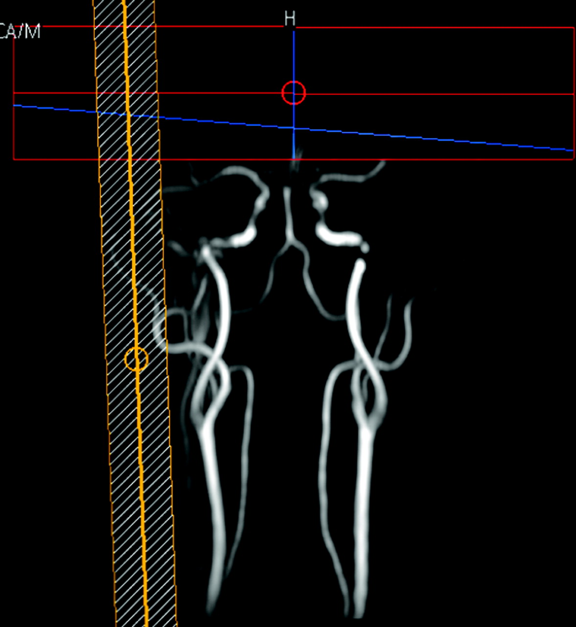
- Fig 1.
On the basis of MR angiography results, we acquired regional perfusion images by placing a selective labeling slab over the external carotid artery.
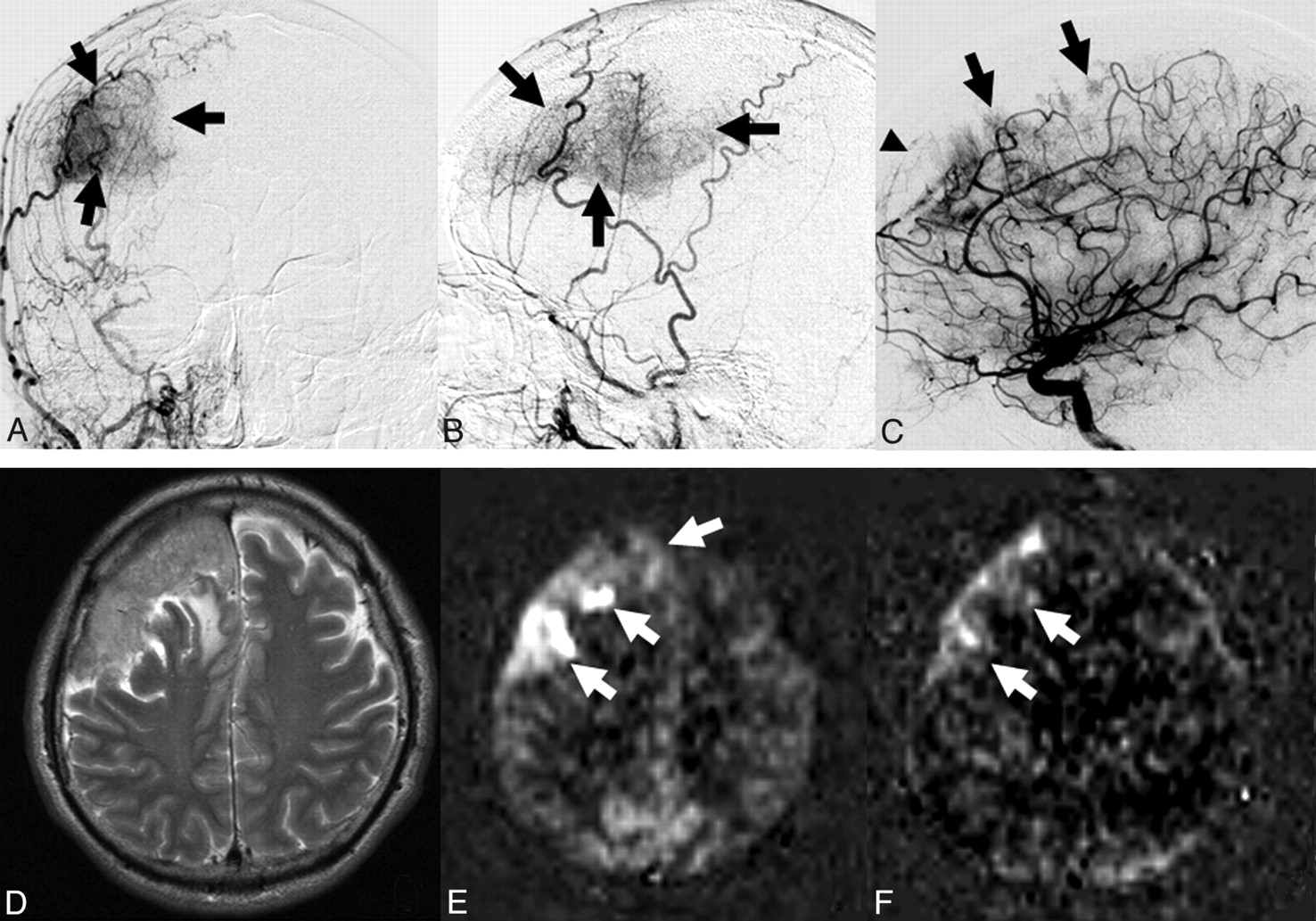
- Fig 2.
A 58-year-old man with malignant meningioma at the convexity (case 4). A and B, Anteroposterior (A) and lateral (B) projections of the right external carotid angiogram show a hypervascular region (arrows) fed by the right middle meningeal artery. C, Lateral projection of the right internal carotid angiogram shows a parasitic supply from the anterior cerebral artery branches (arrows). The tumor is also fed by the falx artery (arrowhead) from the ophthalmic artery. D, T2-weighted image demonstrates a large mass lesion at the right frontal convexity. E, Conventional ASL image shows a vascular territory with higher perfusion than that in the normal-appearing cortex (arrows). The degree of tumor perfusion is classified as grade 3. F, RPI acquired at the same level as E. The extent of the vascular tumor territory is slightly smaller on the RPI than on the conventional ASL image (arrows). The extent of tumor perfusion on the 2 techniques is classified as partially different.
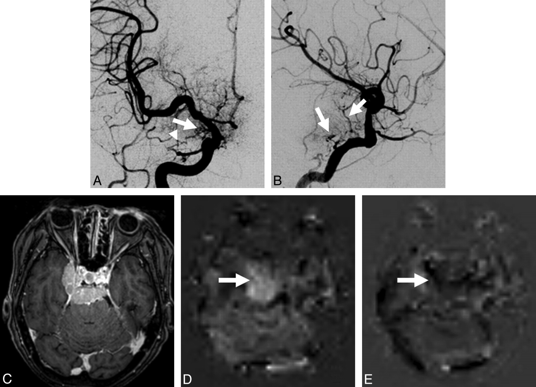
- Fig 3.
A 71-year-old woman with a meningioma at the right cavernous sinus (case 8). A, Anteroposterior projection of the right internal carotid angiogram shows dilated feeding arteries (arrow and arrowhead) from the internal carotid and ophthalmic arteries, respectively. The external carotid artery branches are also seen due to reflux of contrast medium. B, Lateral projection of the left internal carotid angiogram clearly shows dilated feeding arteries (arrows) from the internal carotid artery. C, Contrast-enhanced 3D TFE image demonstrates a well-enhanced mass lesion at the right cavernous sinus and posterior cranial fossa. D, Conventional ASL image shows a tumor vascular territory with higher perfusion than that in the normal-appearing cortex (arrow). The degree of tumor perfusion is classified as grade 3. E, RPI acquired at the same level as D. The hypervascular territory is not depicted on the RPI (arrow). The extent of tumor perfusion on the 2 techniques is classified as completely different.
Tables
- Table 1:
Summary of patient characteristics and image findings in 8 patients with meningioma
Case/Age(yr)/Sex Location/Maximum Diameter (mm) DSAa Rating of Tumor Perfusion on ASL Extent of Tumor Vascular Territory on ASL and RPI Observer 1 Observer 2 Observer 1 Observer 2 1/58/F Posterior fossa/20 + Grade 2b Grade 2b Coincided Coincided 2/73/F Rt middle fossa/40 – Grade 2b Grade 3c Coincided Coincided 3/48/F Rt convexity/55 + Grade 3c Grade 3c Coincided Coincided 4/58/Md Rt convexity/60 + Grade 3c Grade 3c PD PD 5/64/M Lt convexity/45 – Grade 3c Grade 3c Coincided PD 6/58/F Lt middle fossa/33 – Grade 3c Grade 3c PD PD 7/42/M Lt middle fossa/65 + Grade 3c Grade 3c PD PD 8/71/F Rt cavernous sinus/50 + Grade 3c Grade 3c CD CD a + indicates performed; –, not performed.
b Tumor perfusion equivalent to that in the normal-appearing cortex.
c Tumor perfusion higher than that of the normal-appearing cortex.
d Malignant meningioma.
Case/Age(yr)/Sex Location/Maximal Diameter (mm) Feeding Arteries on DSA Extent of Tumor Vascular Territory on ASL and RPI Observer 1 Observer 2 1/58/F Posterior fossa/20 Rt occipital artery Coincided Coincided 3/48/F Rt convexity/55 Rt MMA Coincided Coincided 4/58/Ma Rt convexity/60 Rt MMA, falx artery, parasitic supply from ACA PD PD 7/42/M Lt middle fossa/65 Lt APA, Lt MMA, Lt occipital artery, Rt ICA, parasitic supply from PCA PD PD 8/71/F Rt cavernous sinus/50 Rt ICA, Rt ophthalmic artery CD CD a Malignant meningioma.


