
Article Figures & Data
Figures
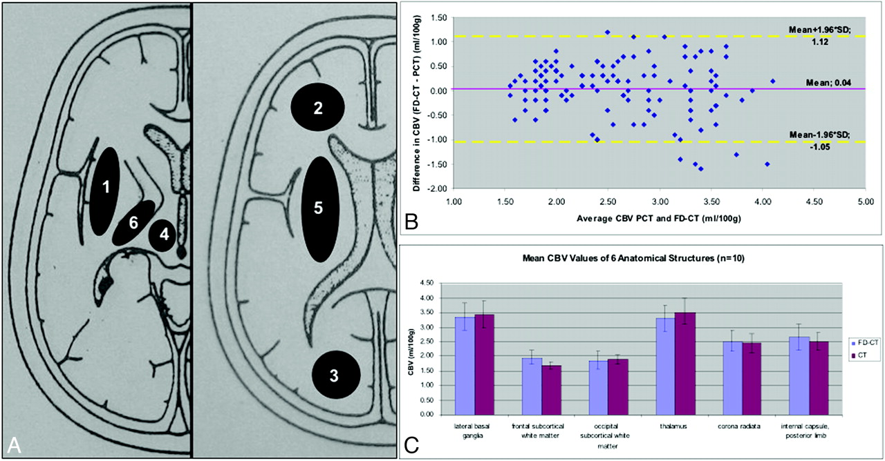
- Fig 1.
A, Regions of interest for CBV measurements: 1) lateral basal ganglia; 2) frontal subcortical white matter; 3) occipital subcortical white matter; 4) thalamus; 5) corona radiata; 6) internal capsule, posterior limb. B, Bland-Altman plot displays slightly higher FD-CBV values (0.04 mL/100 mL) in comparison with CTP. C, Mean CBV values and SDs of the 6 regions of interest show only minimal deviations.
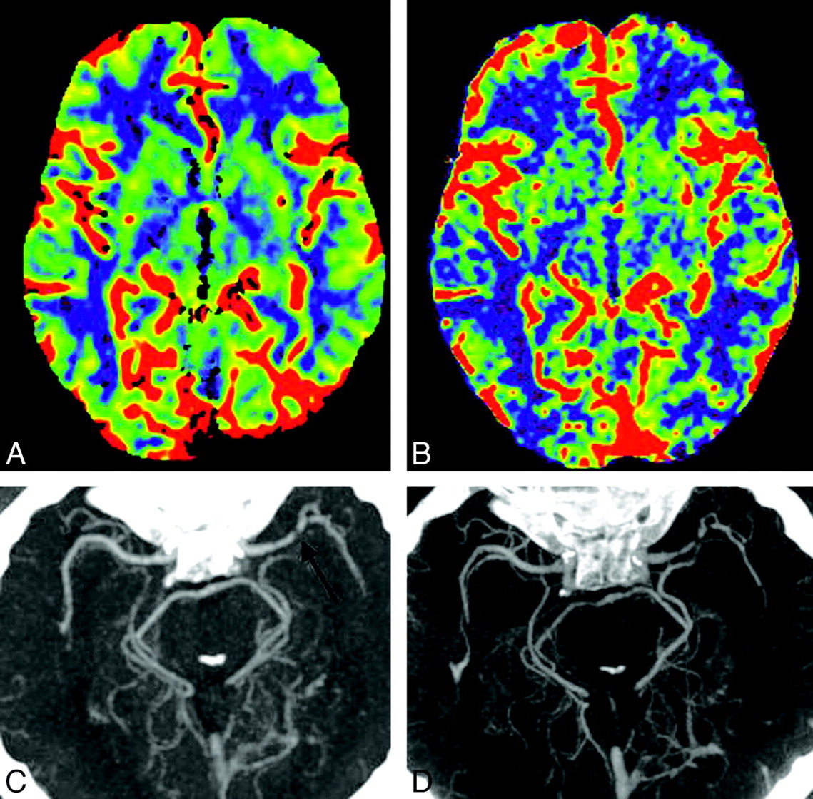
- Fig 2.
CT (A) and FD-CT (B) show the good correlation of CBV color maps. CTA (C) and FD-CTA (D) display a high-grade stenosis (black arrows) of the left MCA (patient 4). Note the perfect delineation of this high-grade stenosis in FD-CTA.
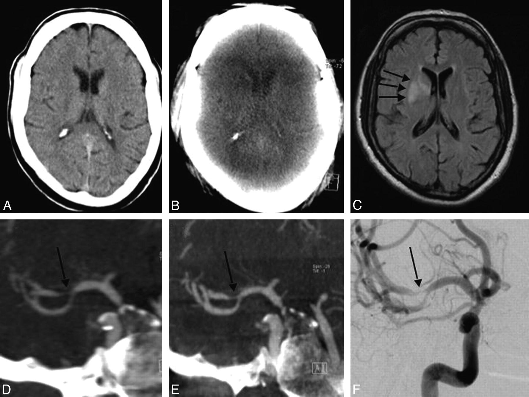
- Fig 3.
A, At initial scanning, the brain appears without any signs of stroke. B, FD-CT performed 24 hours later cannot visualize the stroke region. C, MR imaging also performed 24 hours later confirms infarct demarcation (arrows). D−F, MIP reconstruction of the CTA (D) and FD-CTA (E) corresponds perfectly with the DSA finding (F) of stenosis (arrow) in the distal M1 as well as the proximal M2 segments. Notice the sharp delineation of the vessels in FD-CTA in contrast to that in CTA.
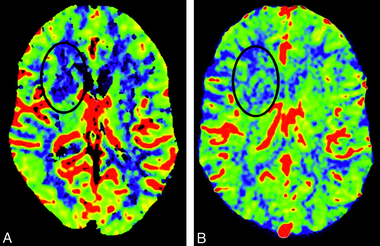
- Fig 4.
A and B, Comparison of CTP-CBV (A) with FD-CBV (B) demonstrates decrease of CBV in the region of the internal capsule, anterior limb. C, The region matches nicely with the MR image (Fig 3C).


Tables
Summary of clinical and imaging characteristics
No. Age (yr) Vascular Pathology Symptoms Treatment CBV Lesion FD-CT CBV CTP-CBV Evans Index Stenosis Grade (%) Interval, CT to FD-CT (hr) FD-CT MSCT FD-CT MSCT DSA 1 81 VA stenosis, right TIA Medical 0.27 0.28 67 66 72 20.5 2 70 Extracranial ICA stenosis TIA Carotid stenting 0.26 0.25 17.75 3 76 MCA occlusion, left Minor stroke Medical Internal capsule, anterior limb 2.6 mL/100 mL 2.7 mL/100 mL 0.31 0.32 18 4 61 MCA stenosis, left TIA Stent 0.42 0.43 94 92 95 16.5 5 61 VA stenosis, left TIA Medical 0.37 0.36 82 78 80 18.5 6 69 VA stenosis, right TIA Medical 0.3 0.31 88 85 84 13.5 7 50 MCA stenosis, right Minor stroke Stent Internal capsule, nterior limb 2.1 mL/100 mL 1.8 mL/100 mL 0.25 0.26 80 81 76 13.75 8 52 MCA occlusion, right Minor stroke Medical Temporal lobe 1.6 mL/100 mL Not visible 0.21 0.22 16.25 9 67 MCA aneurysm TIA Medical 0.28 0.27 14.5 10 67 Carotid aneurysm TIA Medical 0.24 0.25 21.5
In this issue


Jump to section
Related Articles
Cited By...
- Value of Immediate Flat Panel Perfusion Imaging after Endovascular Therapy (AFTERMATH): A Proof of Concept Study
- Clinical Applications of Conebeam CTP Imaging in Cerebral Disease: A Systematic Review
- Dual-Layer Detector Cone-Beam CT Angiography for Stroke Assessment: First-in-Human Results (the Next Generation X-ray Imaging System Trial)
- Dual-Layer Detector Cone-Beam CT Angiography for Stroke Assessment: First-in-Human Results (the Next Generation X-ray Imaging System Trial)
- Dual-Layer Detector Cone-Beam CT Angiography for Stroke Assessment: First-in-Human Results (the Next Generation X-ray Imaging System Trial)
- Distal Vessel Imaging via Intra-arterial Flat Panel Detector CTA during Mechanical Thrombectomy
- Flat-detector computed tomography PBV map in the evaluation of presurgical embolization for hypervascular brain tumors
- Time-Resolved C-Arm Computed Tomographic Angiography Derived From Computed Tomographic Perfusion Acquisition: New Capability for One-Stop-Shop Acute Ischemic Stroke Treatment in the Angiosuite
- Dynamic Angiography and Perfusion Imaging Using Flat Detector CT in the Angiography Suite: A Pilot Study in Patients with Acute Middle Cerebral Artery Occlusions
- Minimally invasive cone beam CT-guided evacuation of parenchymal and ventricular hemorrhage using the Apollo system: proof of concept in a cadaver model
- Exploring the Value of Using Color-Coded Quantitative DSA Evaluation on Bilateral Common Carotid Arteries in Predicting the Reliability of Intra-Ascending Aorta Flat Detector CT-CBV Maps
- Quantitative analysis of high-resolution, contrast-enhanced, cone-beam CT for the detection of intracranial in-stent hyperplasia
- A prospective, multicenter pilot study investigating the utility of flat detector derived parenchymal blood volume maps to estimate cerebral blood volume in stroke patients
- Radiation Doses of Cerebral Blood Volume Measurements Using C-Arm CT: A Phantom Study
- Predictive value of flat-panel CT for haemorrhagic transformations in patients with acute stroke treated with thrombectomy
- C-Arm CT Measurement of Cerebral Blood Volume and Cerebral Blood Flow Using a Novel High-Speed Acquisition and a Single Intravenous Contrast Injection
- Subarachnoid Hyperattenuation on Flat Panel Detector-Based Conebeam CT Immediately after Uneventful Coil Embolization of Unruptured Intracranial Aneurysms
- Frameless multimodal image guidance of localized convection-enhanced delivery of therapeutics in the brain
- Feasibility of Intravenous Flat Panel Detector CT Angiography for Intracranial Arterial Stenosis
- Initial experience with a combined multidetector CT and biplane digital subtraction angiography suite with a single interactive table for the diagnosis and treatment of neurovascular disease
- Monitoring Peri-Therapeutic Cerebral Circulation Time: A Feasibility Study Using Color-Coded Quantitative DSA in Patients with Steno-Occlusive Arterial Disease
- C-Arm CT Measurement of Cerebral Blood Volume Using Intra-Arterial Injection of Contrast Medium: An Experimental Study in Canines
- Feasibility of Cerebral Blood Volume Mapping by Flat Panel Detector CT in the Angiography Suite: First Experience in Patients with Acute Middle Cerebral Artery Occlusions
- Quantitative Evaluation of C-Arm CT Cerebral Blood Volume in a Canine Model of Ischemic Stroke
- Applicability of Tableside Flat Panel Detector CT Parenchymal Cerebral Blood Volume Measurement in Neurovascular Interventions: Preliminary Clinical Experience
- Flat Panel Detector CT, CT Angiography, and CT Perfusion in Stroke