Advertisement

Main menu

  • Home
  • Content
    • Current Issue
    • Publication Preview--Ahead of Print
    • Past Issue Archive
    • Case of the Week Archive
    • Classic Case Archive
    • Case of the Month Archive
    • COVID-19 Content and Resources
  • For Authors
  • About Us
    • About AJNR
    • Editors
    • American Society of Neuroradiology
  • Submit a Manuscript
  • Podcasts
    • Subscribe on iTunes
    • Subscribe on Stitcher
  • More
    • Subscribers
    • Permissions
    • Advertisers
    • Alerts
    • Feedback
  • Other Publications
    • ajnr

User menu

  • Subscribe
  • Alerts
  • Log in

Search

  • Advanced search
American Journal of Neuroradiology

American Journal of Neuroradiology

  • Subscribe
  • Alerts
  • Log in

Advanced Search

  • Home
  • Content
    • Current Issue
    • Publication Preview--Ahead of Print
    • Past Issue Archive
    • Case of the Week Archive
    • Classic Case Archive
    • Case of the Month Archive
    • COVID-19 Content and Resources
  • For Authors
  • About Us
    • About AJNR
    • Editors
    • American Society of Neuroradiology
  • Submit a Manuscript
  • Podcasts
    • Subscribe on iTunes
    • Subscribe on Stitcher
  • More
    • Subscribers
    • Permissions
    • Advertisers
    • Alerts
    • Feedback
  • Follow AJNR on Twitter
  • Visit AJNR on Facebook
  • Follow AJNR on Instagram
  • Join AJNR on LinkedIn
  • RSS Feeds
Research ArticlePharmacology Vignette

Natalizumab (Tysabri)

D.T. Selewski, G.V. Shah, B.M. Segal, P.A. Rajdev and S.K. Mukherji
American Journal of Neuroradiology October 2010, 31 (9) 1588-1590; DOI: https://doi.org/10.3174/ajnr.A2226
D.T. Selewski
  • Find this author on Google Scholar
  • Find this author on PubMed
  • Search for this author on this site
G.V. Shah
  • Find this author on Google Scholar
  • Find this author on PubMed
  • Search for this author on this site
B.M. Segal
  • Find this author on Google Scholar
  • Find this author on PubMed
  • Search for this author on this site
P.A. Rajdev
  • Find this author on Google Scholar
  • Find this author on PubMed
  • Search for this author on this site
S.K. Mukherji
  • Find this author on Google Scholar
  • Find this author on PubMed
  • Search for this author on this site
  • Article
  • Figures & Data
  • Info & Metrics
  • References
  • PDF
Loading

Article Figures & Data

Figures

  • Fig 1.
    • Download figure
    • Open in new tab
    • Download powerpoint
    Fig 1.

    The normal process of leukocyte migration out of blood vessels into tissue involves interactions between leukocytes and endothelial cells including rolling (A), adhesion (B), and extravasation (C). The adhesion molecules α4β1 and α4β7 found on leukocytes are integral in the adhesion process to endothelial cells.
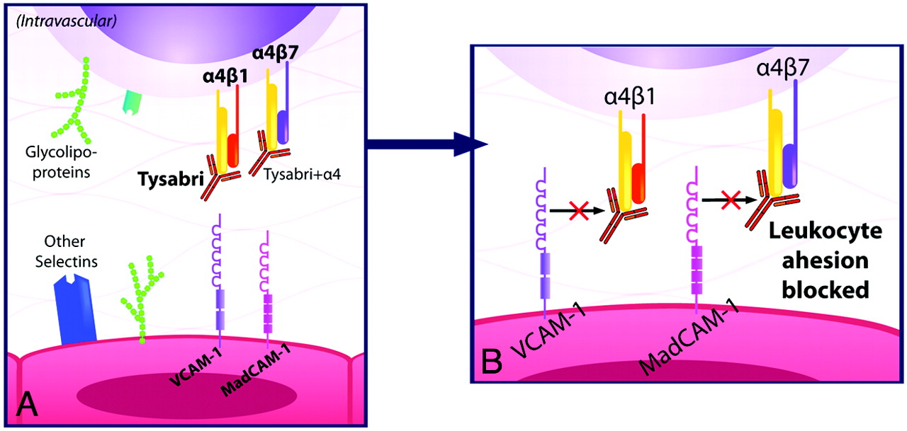
  • Fig 2.
    • Download figure
    • Open in new tab
    • Download powerpoint
    Fig 2.

    A, Natalizumab blocks the adhesion of leukocytes to endothelial cells by blocking the interaction of the α4-integrin subunit of α4β1 with VCAM-1 and of α4β7 with mucosal MAdCAM-1. B, This prevents autoreactive leukocytes from exiting blood vessels and entering target organs to cause inflammation.

  • Fig 3.
    • Download figure
    • Open in new tab
    • Download powerpoint
    Fig 3.

    A 27-year-old man presented with numbness and weakness of both upper extremities and the left lower extremity, with multiple enhancing MR imaging lesions. He was prescribed high-dose β interferon (Rebif) soon after his initial clinical exacerbation but was switched to glatiramer acetate 1 year later due to breakthrough radiologic disease activity. The patient developed new right-sided paresthesias 3 years after his initial presentation. The MR imaging in this figure was performed when the new symptoms developed. A, FLAIR-weighted sagittal FSE image of the brain shows patchy high-signal-intensity areas involving the corpus callosum, brain stem structures, and cerebellum. B, FLAIR-weighted axial FSE image shows multiple patchy areas of high FLAIR signal intensity involving the corpus callosum and bilateral periventricular white matter with the presence of edema around a large right periatrial lesion. C, Postcontrast T1-weighted axial FSE image shows that a majority of the larger lesions exhibit intense patchy enhancement, suggestive of active demyelination.

  • Fig 4.
    • Download figure
    • Open in new tab
    • Download powerpoint
    Fig 4.

    Within 1 month of the MR imaging shown in Fig 3, the patient was started on a course of natalizumab (Tysabri), 300 mg administered intravenously every 4 weeks. Repeat MR imaging after 6 months was performed. A, FLAIR-weighted sagittal FSE image of the brain shows improvement in the patchy high-signal-intensity area of the corpus callosum with resolution of lesions involving the brain stem structures and cerebellum. B, FLAIR-weighted axial FSE image shows marked improvement in the areas of demyelination involving the corpus callosum and bilateral periventricular white matter. C, Postcontrast T1-weighted axial FSE image shows only 1 small area of enhancement in the right periventricular white matter, with lack of enhancement of the rest of the enhancing lesions.

PreviousNext
Back to top

In this issue

American Journal of Neuroradiology: 31 (9)
American Journal of Neuroradiology
Vol. 31, Issue 9
1 Oct 2010
  • Table of Contents
  • Index by author
Advertisement
Print
Download PDF
Email Article

Thank you for your interest in spreading the word on American Journal of Neuroradiology.

NOTE: We only request your email address so that the person you are recommending the page to knows that you wanted them to see it, and that it is not junk mail. We do not capture any email address.

Enter multiple addresses on separate lines or separate them with commas.
Natalizumab (Tysabri)
(Your Name) has sent you a message from American Journal of Neuroradiology
(Your Name) thought you would like to see the American Journal of Neuroradiology web site.
CAPTCHA
This question is for testing whether or not you are a human visitor and to prevent automated spam submissions.
Citation Tools
Natalizumab (Tysabri)
D.T. Selewski, G.V. Shah, B.M. Segal, P.A. Rajdev, S.K. Mukherji
American Journal of Neuroradiology Oct 2010, 31 (9) 1588-1590; DOI: 10.3174/ajnr.A2226

Citation Manager Formats

  • BibTeX
  • Bookends
  • EasyBib
  • EndNote (tagged)
  • EndNote 8 (xml)
  • Medlars
  • Mendeley
  • Papers
  • RefWorks Tagged
  • Ref Manager
  • RIS
  • Zotero
Share
Natalizumab (Tysabri)
D.T. Selewski, G.V. Shah, B.M. Segal, P.A. Rajdev, S.K. Mukherji
American Journal of Neuroradiology Oct 2010, 31 (9) 1588-1590; DOI: 10.3174/ajnr.A2226
del.icio.us logo Digg logo Reddit logo Twitter logo CiteULike logo Facebook logo Google logo Mendeley logo
  • Tweet Widget
  • Facebook Like
  • Google Plus One
Purchase

Jump to section

  • Article
    • Abstract
    • Abbreviations
    • References
  • Figures & Data
  • Info & Metrics
  • References
  • PDF

Related Articles

  • No related articles found.
  • PubMed
  • Google Scholar

Cited By...

  • Encephalitogenic and Regulatory CD8 T Cells in Multiple Sclerosis and Its Animal Models
  • Crossref
  • Google Scholar

This article has not yet been cited by articles in journals that are participating in Crossref Cited-by Linking.

More in this TOC Section

  • Dabigatran (Pradaxa)
  • Clopidogrel (Plavix)
  • Trastuzumab (Herceptin)
Show more Pharmacology Vignette

Similar Articles

Advertisement

News and Updates

  • Lucien Levy Best Research Article Award
  • Thanks to our 2022 Distinguished Reviewers
  • Press Releases

Resources

  • Evidence-Based Medicine Level Guide
  • How to Participate in a Tweet Chat
  • AJNR Podcast Archive
  • Ideas for Publicizing Your Research
  • Librarian Resources
  • Terms and Conditions

Opportunities

  • Share Your Art in Perspectives
  • Get Peer Review Credit from Publons
  • Moderate a Tweet Chat

American Society of Neuroradiology

  • Neurographics
  • ASNR Annual Meeting
  • Fellowship Portal
  • Position Statements

© 2023 by the American Society of Neuroradiology | Print ISSN: 0195-6108 Online ISSN: 1936-959X

Powered by HighWire