
Abstract
BACKGROUND AND PURPOSE: The PVICC of the craniospinal compartment defines the shape of the pressure-volume curve and determines the damping of cyclic arterial pulsations. Despite no reports of direct measurements of the PVICC among healthy elderly, it is believed that a change away from adequate accommodation of cardiac-related pulsations may be a pathophysiologic mechanism seen in neurodegenerative disorders such as Alzheimer disease and idiopathic normal pressure hydrocephalus. In this study, blood and CSF flow measurements are combined with lumbar CSF infusion measurements to assess the craniospinal PVICC and its distribution of cranial and spinal compartments in healthy elderly.
MATERIALS AND METHODS: Thirty-seven healthy elderly were included (60–82 years of age). The cyclic arterial volume change and the resulting shift of CSF to the spinal compartment were quantified by PC-MR imaging. In addition, each subject underwent a lumbar CSF infusion test in which the magnitude of cardiac-related pulsations in intracranial pressure was quantified. Finally, the PVI was calculated by using a mathematic model.
RESULTS: After excluding 2 extreme values, the craniospinal PVICC was calculated to a mean of 9.8 ± 2.7 mL and the estimated average 95% confidence interval of individual measurements was ± 9%. The average intracranial and spinal contributions to the overall compliance were 65% and 35% respectively (n = 35).
CONCLUSIONS: Combining lumbar CSF infusion and PC-MR imaging proved feasible and robust for assessment of the craniospinal PVICC. This study produced normative values and showed that the major compensatory contribution was located intracranially.
Abbreviations
- ECG
- electrocardiogram
- ΔICP
- intracranial pulse pressure magnitude
- ΔPVICC
- relative width of the 95% confidence interval of the calculated PVICC
- ΔRPPC
- width of the 95% confidence interval of the calculated RPPC
- ΔVART
- arterial volume change
- ΔVbolus
- volume infused in a lumbar CSF bolus infusion test
- ΔVIC
- volume accommodated by the intracranial compartment
- ΔVSC
- volume displaced to the spinal compartment
- ICP
- intracranial pressure
- ICPend
- ICP after lumbar CSF bolus infusion test
- ICPstart
- ICP before lumbar CSF bolus infusion test
- P1 and P0
- pressure constants
- PC-MR imaging
- phase contrast MR imaging
- PVI
- pressure-volume index
- PVIbolus
- PVI of the craniospinal cavity estimated from a lumbar CSF bolus infusion test
- PVICC
- PVI of the craniospinal cavity
- PVIIC
- PVI of the intracranial compartment
- PVISC
- PVI of the spinal compartment
- RPPC
- relative pulse-pressure coefficient
PVICC is a measure of compliance of the craniospinal cavity, defined as the amount of volume that has to be added to raise the ICP 10 times.1 In neurointensive care, PVICC is an important parameter defining the shape of the pressure-volume curve. The concept of compliance and PVICC can also be applied to arterial pulsations. A bolus of blood enters the craniospinal cavity during each systole. A decreased PVICC corresponds to a reduced ability to accommodate the pulsations generated by these boluses and could, with time, result in clinical symptoms. It has been hypothesized that a mechanical dysfunction of this type has implications for Alzheimer disease and vascular dementia.2,3 Moreover, increased cerebrovascular pulsatility has been linked to the development of white matter lesions, and elevated ICP pulse amplitude has been shown to be important in predicting outcome following shunt surgery in idiopathic normal pressure hydrocephalus.4,5 Together these observations illuminate how alterations in arterial pulsations (often associated with increasing age) and pulsation dampening may be involved in several degenerative neurologic disorders among the elderly.
The relationship between ICP and volume has empirically been shown as follows6,7:
 where VCC is the volume of the system. Repeated bolus testing (ie, a lumbar puncture followed by controlled infusions of mock CSF and analysis of the corresponding ICP responses) is a common method to assess PVICC. Unfortunately little is known about PVICC healthy individuals. The PVICC is the sum of compensatory mechanisms in the cranial and spinal compartments. Animal experiments have produced quantifications of the compliance of each compartment.8 These methods require a physical separation between the compartments and have not been applied to human subjects. In general, lumbar CSF infusion tests cannot provide information on how the compliance is distributed within the craniospinal cavity. By measuring flow of blood and CSF using flow-sensitive PC-MR imaging, it is possible to quantify both the pulsating blood flow to the brain and the resulting CSF volume shift to the spinal compartment that occurs naturally within every cardiac cycle.9 PC-MR imaging alone has been used to derive parameters related to compliance, but without a direct measurement of ICP, the PVICC cannot be explicitly calculated (equation 1).9,10 In this study, we propose and evaluate a combination of lumbar CSF infusion tests and PC-MR imaging flow measurements to assess PVICC and its distribution between the intracranial and spinal compartments. We use this method to describe the normal compensatory capacity in a group of healthy elderly.
Materials and Methods
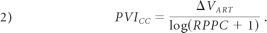
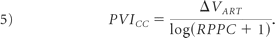
A property of the craniospinal system, consistent with the mathematic model of equation 1, is the linear relationship between mean ICP and ΔICP (Fig 1).7 The slope of the linear relationship, denoted RPPC, can be assessed during a lumbar CSF infusion study.11 Assessment of RPPC in combination with a measurement of the ΔVART occurring during a heart cycle enables calculation of PVICC (see Appendix for derivation). The relationship can be written as
 Furthermore, assuming a communicating system without any differences between RPPC measured intracranially and in the spinal compartment enables calculation of the compliances of the intracranial and spinal compartments (PVIIC and PVISC, respectively) by replacing ΔVART with the ΔVSC or the ΔVIC in equation 2. ΔVART and ΔVSC can be measured with PC-MR imaging.10,12 Moreover, the difference between the arterial volume increase and the volume shifted to the spinal compartment is the ΔVIC. It follows that PC-MR imaging and lumbar CSF infusion measurements can be combined to calculate PVICC, PVIIC, and PVISC. In this study, initial MR imaging measurements for anatomic assessments of the brain were conducted, immediately followed by flow quantification with PC-MR imaging (Fig 2A). If no contraindication was revealed from the MR imaging investigation, the subject continued to the lumbar CSF infusion investigation (Fig 2B). The output from the PC-MR imaging measurements and the lumbar CSF infusion investigation was combined by using equation 2 (Fig 2C).

A subject recording of ΔICP and ICP from a constant pressure lumbar CSF infusion test. The RPPC is the slope of the linear regression line between ΔICP and ICP. P0 is calculated as the pressure at which this line intersects with ΔICP = 0.

An overview of how the infusion and MR imaging modalities are combined to estimate compliance indices.
Subjects
An ad was put in the local paper inviting healthy volunteers (60–82 years of age) to apply for a research project regarding MR imaging and lumbar puncture (including a lumbar CSF dynamic investigation). Of the 149 that answered the ad, 59 persons were called for an interview, including a neurologic examination. Fifty subjects qualified. All had a Mini-Mental State Examination score of >28 points, and none had >2 of the vascular risk factors, smoking, hypertension, or hyperlipidemia.13 Advanced vascular diseases (eg, diabetes, previous stroke, or myocardial infarction) were exclusion criteria, as well as use of anticoagulants, benzodiazepines, or antidepressants. Five of the volunteers were excluded after the MR imaging, and the lumbar CSF infusion investigations could not be carried out in 4 individuals. Two MR imaging flow measurements were discarded because of cardiac synchronization difficulties and 2 lumbar CSF infusion investigations could not be used because of needle problems (different subjects). Thus, the studied group consisted of 37 healthy elderly (22 women and 15 men). Fifteen subjects had their lumbar CSF infusion investigation within 1 hour following the MR imaging; 21, the next day; and 1 subject, 6 days after the MR imaging. The mean age was 71 ± 6 years. The local ethics committee approved the study. Informed consent was obtained from all participants.
MR Imaging Measurements
Measurements were performed on a 3T Achieva scanner (Philips Healthcare, Best, the Netherlands). Routine sequences (T1, T2*, T2, and fluid-attenuated inversion recovery) were performed to assess criteria set for inclusion as a healthy subject. The PC-MR imaging sequences had a scan matrix of 128 × 128–160 × 160, a 5- to 6-mm section thickness, a 10- to 16-ms TR, a 6- to 11-ms TE, a flip angle of 10°-15°, and a 2-fold signal-intensity averaging. The velocity sensitization was set to 70 cm/s for blood assessments and 7 cm/s for CSF assessments. Retrospective triggering, ECG or peripheral, was used to synchronize the PC-MR imaging sequence to the cardiac cycle for proper sampling. Thirty-two phases were reconstructed. CSF and blood flows were quantified at the level of the first and second cervical vertebrae (thus proximal to the anterior spinal artery branch). Vessel lumen segmentation was performed manually in ImageJ (http://rsb.info.nih.gov/ij).14 ΔVART was calculated from the sum of the blood flows in the internal carotid and vertebral arteries (Fig 3). The CSF flow at the cervical level was used to estimate ΔVSC. Subtraction of the spinal CSF flow from the summed arterial flow curve was used to calculate ΔVIC.

A, Total cerebral arterial blood flow (summated flow of the internal carotid and vertebral arteries). B, Cumulative integration of the curve in A with the average flow subtracted yields the arterial volume change during a cardiac cycle. ΔVART was defined as the largest volume difference during a cardiac cycle.
Lumbar CSF Infusion Investigation
A fully automatic lumbar CSF infusion apparatus, in which the subject is in a supine position, was used.15 It uses 2 needles inserted in the lumbar canal, 1 for infusion of a Ringer acetate solution and 1 for pressure measurements. Investigations started with a recording of the resting ICP. This was followed by a CSF infusion phase in which the pressure was regulated to 6 predetermined and constant pressure levels, separated by 0.4 kPa (Fig 4). In a few exceptions (n = 4) with partial obstruction of the infusion needle, the operator switched to a pattern with a constant infusion rate of 1.5 mL/min for 20 minutes. The pressure was sampled at 100 Hz. In 26 subjects, the heart rate was monitored by ECG for evaluation of agreement with the heart rate of the MR imaging investigations. There was no significant difference between the heart rate measured during the MR imaging and during the lumbar CSF infusion test (63.9 versus 63.1 beats per minute; mean difference, 0.8 beats per minute; paired t test, P = .5). To calculate RPPC, we estimated a ΔICP by calculating a pulse pressure as the difference between the maximum and minimum ICP in 1.5-second time windows after applying a forward-backward fifth-order high-pass Butterworth filter with a cutoff frequency of 0.5 Hz. For all pressure levels of the constant pressure infusion pattern, including baseline, the ΔICP was defined as the median pressure pulse. The RPPC was calculated by least squares linear regression between the ΔICP and median ICP of the different pressure levels. P0 was determined as the crossing between the regression line and the pressure axis (Fig 1). In the case of a constant infusion, ΔICP and average ICP for all 1.5-second intervals of the infusion phase were used in the regression. The uncertainty of the RPPC parameter, denoted ΔRPPC, was approximated for each investigation by calculating the width of the 95% confidence interval from the regression analysis.

An ICP recording from a lumbar CSF infusion test. A, Baseline ICP recording. B, Infusion to predetermined ICP levels. C, Relaxation phase allowing ICP to normalize. D, A bolus test (rapid infusion of a 5.6-mL artificial CSF).
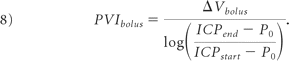
At the end of the investigation, 32 of the subjects were additionally investigated with a single bolus infusion (Fig 4), in which a 5.6-mL-volume ΔVbolus was rapidly administered while the pressure response was recorded.1,16 Equation 8 in the Appendix was used for calculating the PVIbolus.17 The ICPstart, and ICPend were measured as the average ICP during the 10-second periods directly before the start and immediately after the end of the bolus infusion phase. A P0 from the RPPC slope of the infusion test performed before the bolus test was used. ΔPVIcc was calculated by using propagation of error analysis applied on equation 2, together with ΔRPPC as described above and with an assumed precision in the volume displacement estimation of an ΔVART of ±0.1 mL.
Results
From the PC-MR imaging investigation, ΔVART was measured to a mean of 1.98 ± 0.43 mL, and ΔVIC and ΔVSC were measured to 1.36 ± 0.44 mL and 0.68 ± 0.23 mL, respectively (n = 37). The estimations from the lumbar CSF infusion tests yielded a mean RPPC of 0.62 ± 0.25 with a mean ΔRPPC of ±0.07 (n = 37). By combining the parameters from the lumbar CSF infusion test and the PC-MR imaging investigation, we calculated PVICC to 11.8 ± 9.0 mL, PVIIC to 8.4 ± 8.1 mL, and PVISC to 3.9 ± 2.5 mL (n = 37). Two subjects with extreme PVICC estimations contributed strongly to the variation in PVICC (Fig 5). Without extreme values, PVICC was 9.8 ± 2.7 mL, PVIIC was 6.7 ± 2.6 mL, and PVISC was 3.4 ± 1.2 mL (n = 35). In relative figures, 65% (range, 44%–89%) of the compensating capacity was located intracranially and the remaining 35% (range, 11%–56%) was located spinally (n = 35). The average ΔPVIcc was calculated to ± 9%. The average PVIbolus was calculated to 11.2 ± 6.9 mL (n = 32). Without extreme values, the PVIbolus was 10.2 ± 3.7 mL (n = 30). Although the mean values for PVICC and PVIbolus were similar, the correlation of individual observations was low (Fig 5).

The craniospinal PVI calculated from combined MR imaging and infusion (y-xis) and from the bolus test (x-axis). The difference was not statistically different from zero, P = .83 from a paired t test. There was no significant correlation after excluding 2 extreme values (dashed contours) (P = .8778).
Discussion
A method to combine data collected from PC-MR imaging measurements and lumbar CSF infusion studies was presented. This provided a different analysis of the craniospinal compliance properties than that achieved by analyzing results from the different measurement techniques separately. The methodology only required minor additions to the routine procedure for patients referred for a lumbar CSF infusion study because almost all undergo an MR imaging investigation that may easily include PC-MR images. Estimations of PVICC were obtained from the combined methodology and estimations of PVIbolus from a single bolus test. The average PVICC and average PVIbolus were comparable with previous reports of constant-rate infusion studies in subjects with hydrocephalus but were lower than those in previous reports of bolus investigations performed in admitted neurologic and neurosurgical subjects (Table). This may be an indication of actual differences in PVICC between healthy elderly and other groups of patients. If so, it seems as if healthy subjects had a PVICC in the lower end of the spectrum. This was not expected because the general hypothesis is that PVICC would be higher among the healthy and that a low PVICC is pathologic. However, in the Table, studies using a slightly different pressure-volume model without a constant term account for the reports of high PVICC values,1,18 while those using a pressure-volume model with a constant term (as used in this study) have produced reports of PVICC more similar to that generated in this study.19,20 Aside from differences potentially originating in the choice of model, aging is known to influence the compliance of the craniospinal cavity,20 and thus addition of normative data of elderly subjects is crucial.
Reports of PVICC in various states
The ΔPVICC indicated the robustness of the method. How compliance was distributed between the intracranial and spinal compartments was investigated. To our knowledge, such reports of basic physiology are missing in the literature. In terms of average compliance, 65% was found intracranially and 35% spinally. This observation was in agreement with reported values in cats (68% and 32% intracranially and spinally, respectively).8 This might reflect the fact that compressible intracranial veins were a major compliance in the system. While the intracranial compartment was the predominant contributor to the PVICC, the individual variations of this distribution were large.
Assessment of the cardiac-related pulsatile intracranial dynamics is increasingly used to understand the physiology and pathology of the CSF system.21–23 PVICC together with ΔVCC determines the impact on the brain caused by the arterial pulsations (approximately 30 million each year). With increasing age, arterial physiology changes. Arterial stiffness and windkessel dysfunction of the aorta stresses the microcirculation.24 The cyclic ΔVART increases.3 PVICC is a quantification of how well these increased pulsations are cushioned. Ventricular dilation is associated with cognitive and gait impairment.25 That inadequate absorption of the arterial pulse can promote ventricular dilation is a persistent hypothesis on the etiology regarding the development of communicating hydrocephalus.26,27 Moreover, the growing concept of pulse wave encephalopathy has been attributed to several other disorders (Alzheimer disease, mild cognitive impairment, age-related white matter changes).2,28,29 This mechanism may involve a pathologic combination of altered PVICC with or without increased ΔVART. Because the combined MR imaging and lumbar CSF infusion test quantify both parameters (in a time regime of a cardiac cycle), this approach would be particularly useful in studying the occurrence and nature of pulse wave encephalopathy.
Because measurements of PVICC entirely based on lumbar infusion tests are associated with within-subject systematic errors, a new approach is desirable.19 A challenge with current infusion tests is that induced pressure variations are slow. During a time window much wider than a cardiac cycle, autoregulatory vasogenic volume variations will cause both systematic and randomized errors in the estimated PVICC, a possible explanation of previously reported discrepancies between different infusion patterns within subjects.30
This study was based on several assumptions that deserve specific attention. We assumed that it is possible to separate the MR imaging and infusion measurements in time. This requires the measured parameters of equation 2 to remain unchanged between the measurements. Within limits, RPPC is independent of ICP, which might change between the measurements.7 Furthermore, the pressure-volume model of equation 1 together with the well-determined RPPC (ie, a small ΔRPPC) indicates that ΔVART is, in fact, also independent of ICP. These parameters might, however, be altered by a change in heart rate, affecting the cardiac output between the measurements. In this study, no significant difference between the heart rate measured at the different modalities was observed. The calculations rely on the assumption that RPPC agrees between the intracranial and spinal compartments. This requires a low resistance between the 2 compartments, a likely situation except in spinal stenosis.
The craniospinal system is built up with active components generating volume variations and passive components absorbing these variations. The sum of all passive components will form the compliance of the system. In this study, we have considered 2 types of active components. The first consists of craniospinal arterial vessels. These arterial vessels expand during each cardiac cycle. The second active component is external lumbar CSF infusion. If any of these active volume changes are known, the pressure response they generate can be used to quantify the compliance of the system. In this article, we assumed that the volume change, measured with PC-MR imaging of the internal carotid and vertebral arteries, will be entirely transferred as heart-beat-related craniospinal arterial volume changes. This view is in accordance with having arterial pulsations transmitted through the arterial wall in a way that blood at some point in the capillary network flows without oscillations.31 By measuring blood flow at the cervical level, we assumed that the entire arterial volume increase of the craniospinal system could be calculated. This is a generalization because arterial supply to lower parts of the spinal cord is not included (eg, the anterior spinal artery is included but segmental and radicular arteries are not included). The magnitude of the missing spinal arterial flow is difficult to estimate because there are no reliable reports of this quantity.32 However, this missing flow is likely a small fraction compared with the blood flow to the brain, and thus the resulting underestimation of PVICC is also small.
Regarding the comparison between the new methodology and the bolus test, the average PVICC and average PVIbolus agreed but individual observations did not correlate (Fig 5). A likely explanation is that a single bolus test does not estimate the craniospinal compliance with precision, whereby a series of boluses are a common choice.16,33 In fact, the combined MR imaging and lumbar CSF infusion data to determine PVICC are in analogy with an averaging of hundreds of pressure responses caused by physiologic bolus excitations. Thus, this method potentially generated a more robust and accurate estimate than that of the single external bolus excitation.
In Fig 5, two extreme values were identified. One had a low ICP response from the bolus test together with a very low RPPC (the upper right extreme value), while the other had a normal ICP response during the bolus test but a low RPPC. Without these measurements, the variance in PVICC and PVIbolus was greatly decreased and the reduced dataset was believed to be representative of an elderly population. However, it could not be exclusively deduced if the extreme measurements were physiologic or due to needle problems. Nevertheless, extreme values appear in the measurements, and their origin should be further investigated.
Conclusions
The combination of PC-MR imaging measurements and a lumbar CSF dynamic test with an established mathematic model of the craniospinal system proved feasible and robust for assessing the compensatory mechanisms of the craniospinal system and its intracranial and spinal compartments. This study produced normative PVI values for healthy elderly and showed that the major compensatory contribution is located intracranially.
Appendix
The RPPC slope can be mathematically derived by investigating the pressure response following a change in volume7:
 This can be re-expressed to define RPPC as
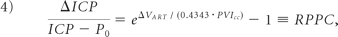 where ΔVart is the cyclic volume displacement related to cardiac pulsations. This can be rewritten to express the PVI as
 Finally, PVIbolus is presented. Starting with equation 3 and by letting the volume displacement represent that of a bolus infusion, we can rewrite the relationship as
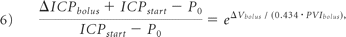 where ICPstart represents the resting pressure preceding the bolus infusion. The pressure response can be expressed as
 where ICPend is the pressure when the bolus volume is administered. It follows that PVIbolus can be expressed as
Footnotes
-
The study was supported by the joint initiative, MTBH, by the Swedish Foundation for Strategic Research, the Swedish Research Council, and the Swedish Governmental Agency for Innovation Systems.
Indicates open access to non-subscribers at www.ajnr.org
References
- Received January 14, 2010.
- Accepted after revision March 26, 2010.
- Copyright © American Society of Neuroradiology
In this issue


Jump to section
Related Articles
Cited By...
- Modeling CSF circulation and the glymphatic system during infusion using subject specific intracranial pressures and brain geometries
- Human intracranial pulsatility during the cardiac cycle: a computational modelling framework
- Spinal Compliance Curves: Preliminary Experience with a New Tool for Evaluating Suspected CSF Venous Fistulas on CT Myelography in Patients with Spontaneous Intracranial Hypotension
- Measuring Pulsatile Flow in Cerebral Arteries Using 4D Phase-Contrast MR Imaging
- Pulsatility in CSF dynamics: pathophysiology of idiopathic normal pressure hydrocephalus
- Blood Flow of Ophthalmic Artery in Healthy Individuals Determined by Phase-Contrast Magnetic Resonance Imaging