
Article Figures & Data
Figures
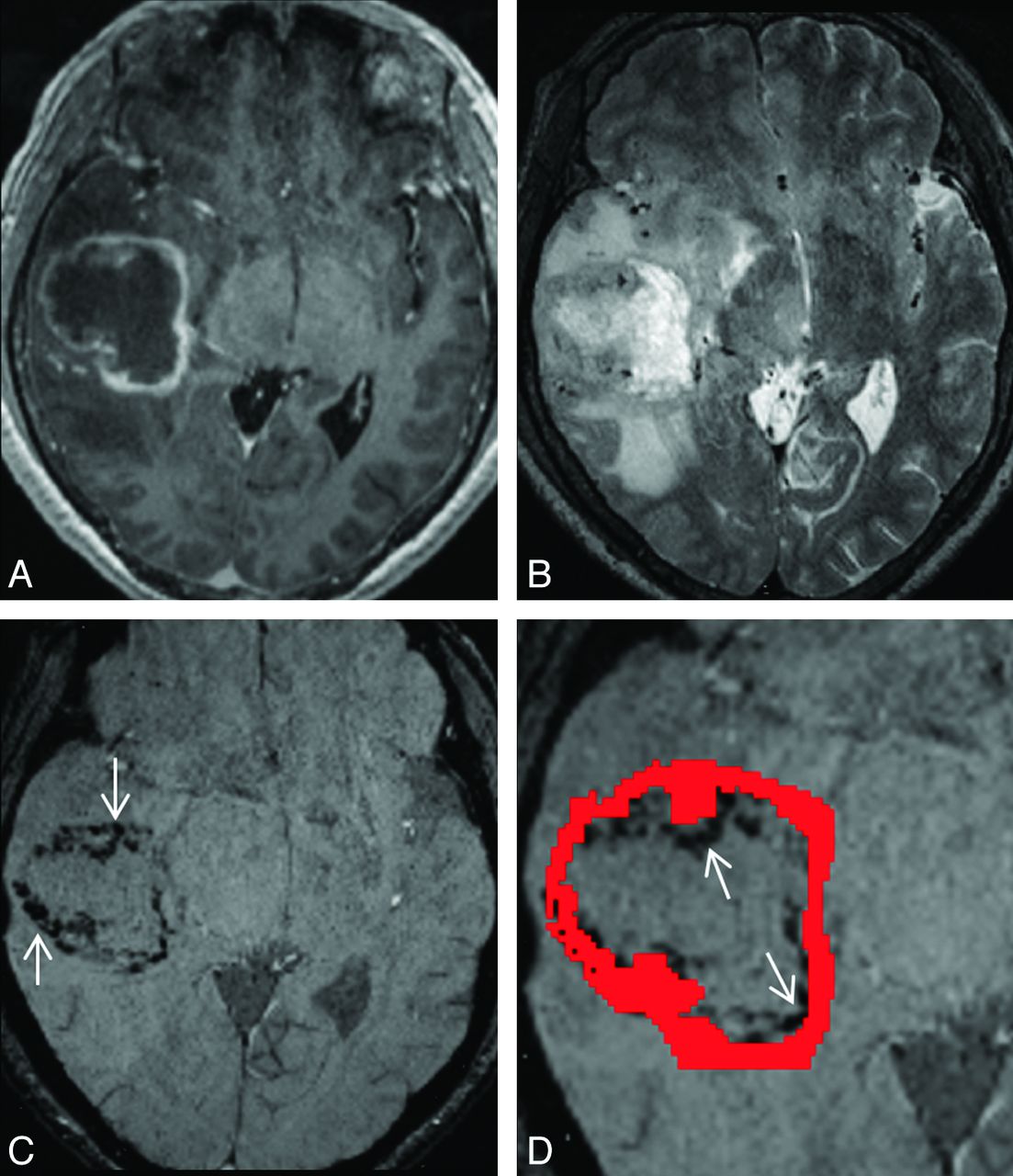
- Fig 1.
A 56-year-old woman with 2 right occipital pyogenic brain abscesses. A, Transverse contrast-enhanced MPRAGE shows 2 adjoining rim-enhancing masses in the right occipital lobe. B, On the transverse T2-weighted image, an incomplete hypointense rim (arrow) is present at the lesion margin. C, On transverse SWI, the abscesses are bordered by 2 concentric rims, with the outer one being hypointense (arrow), and the inner one, hyperintense (arrowhead) relative to cavity content, forming the dual rim sign. D, On the close-up view of the SWI with the contrast-enhancing rim (red region of interest) overlaid, the hyperintense rim (arrowhead) is inner to the contrast-enhancing rim.
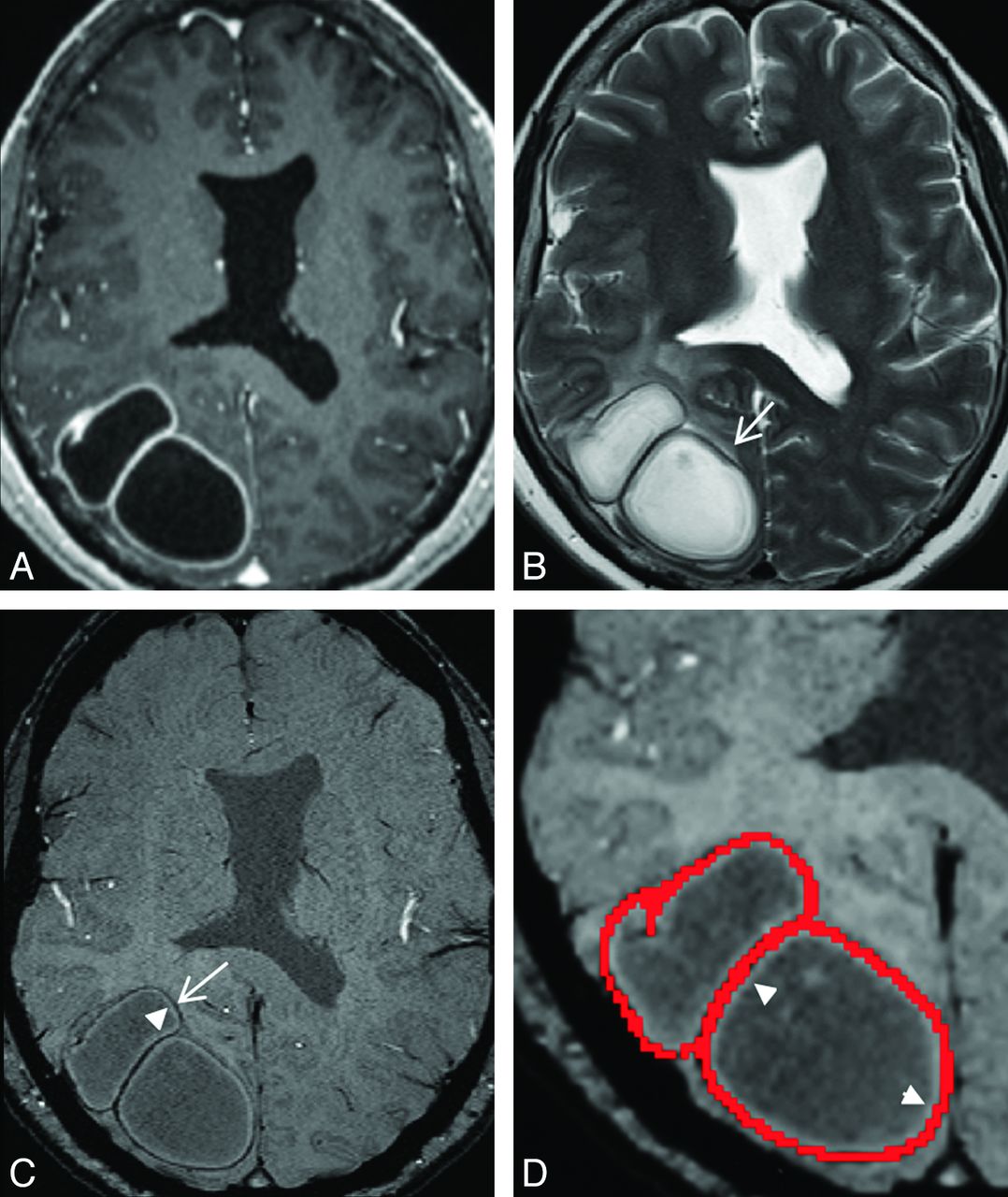
- Fig 2.
A 57-year-old man with a right temporal necrotic glioblastoma. A, Transverse contrast-enhanced MPRAGE shows a rim-enhancing mass in the right temporal lobe. B, On the transverse T2-weighted image, the hypointense rim is absent. C, On the transverse SWI, abundant hypointensities (arrows) with an irregular contour forming an incomplete rim are present at the lesion margin. D, On the close-up view of SWI with a contrast-enhancing rim (red region of interest) overlaid, some of these hypointensities (arrows) are inner to the contrast-enhancing rim, while some are overlapping the contrast-enhancing rim.
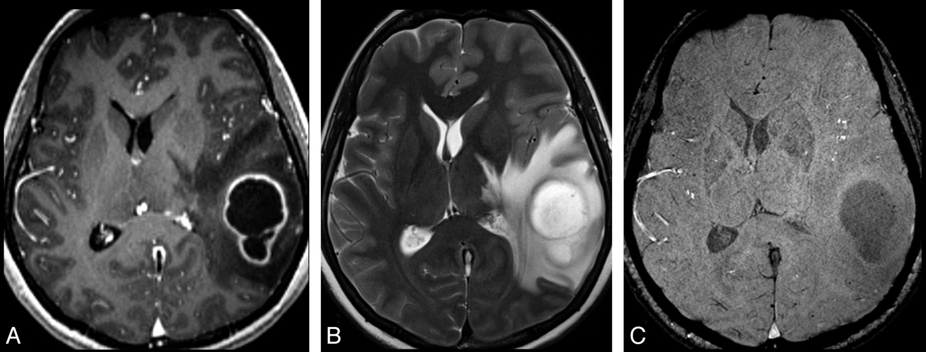
- Fig 3.
A 40-year-old woman with a left temporal necrotic glioblastoma. A, Transverse contrast-enhanced MPRAGE shows a rim-enhancing mass in the left temporal lobe. The hypointense rim is absent on both transverse T2W (B) and SW (C) images.

Tables
Comparisons of the imaging features of the lesion margins on SWI between abscesses and glioblastomas
Features Abscess GB P Value SEN SPE Accuracy PPV NPV Hypointense rim Prevalence 0.059 Absent 0 5 Present 12 15 Border <.001 91.7 86.7 88.9 84.6 92.9 Incomplete 1 13 Completea 11 2 Contour <.001 75.0 86.7 81.5 81.8 81.3 Smootha 9 2 Irregular 3 13 Locationb <.001 91.7 86.7 88.9 84.6 92.9 Inner 0 9 Samea 11 2 Inner + Same 1 4 Dual rim sign <.001 75.0 100 90.6 100 87.0 Absent 3 20 Presenta 9 0


