Advertisement

Main menu

  • Home
  • Content
    • Current Issue
    • Publication Preview--Ahead of Print
    • Past Issue Archive
    • Case of the Week Archive
    • Classic Case Archive
    • Case of the Month Archive
    • COVID-19 Content and Resources
  • For Authors
  • About Us
    • About AJNR
    • Editors
    • American Society of Neuroradiology
  • Submit a Manuscript
  • Podcasts
    • Subscribe on iTunes
    • Subscribe on Stitcher
  • More
    • Subscribers
    • Permissions
    • Advertisers
    • Alerts
    • Feedback
  • Other Publications
    • ajnr

User menu

  • Subscribe
  • Alerts
  • Log in

Search

  • Advanced search
American Journal of Neuroradiology

American Journal of Neuroradiology

  • Subscribe
  • Alerts
  • Log in

Advanced Search

  • Home
  • Content
    • Current Issue
    • Publication Preview--Ahead of Print
    • Past Issue Archive
    • Case of the Week Archive
    • Classic Case Archive
    • Case of the Month Archive
    • COVID-19 Content and Resources
  • For Authors
  • About Us
    • About AJNR
    • Editors
    • American Society of Neuroradiology
  • Submit a Manuscript
  • Podcasts
    • Subscribe on iTunes
    • Subscribe on Stitcher
  • More
    • Subscribers
    • Permissions
    • Advertisers
    • Alerts
    • Feedback
  • Follow AJNR on Twitter
  • Visit AJNR on Facebook
  • Follow AJNR on Instagram
  • Join AJNR on LinkedIn
  • RSS Feeds
Research ArticleExtracranial Vascular
Open Access

Predicting Carotid Plaque Characteristics Using Quantitative Color-Coded T1-Weighted MR Plaque Imaging: Correlation with Carotid Endarterectomy Specimens

S. Narumi, M. Sasaki, H. Ohba, K. Ogasawara, M. Kobayashi, T. Natori, J. Hitomi, H. Itagaki, T. Takahashi and Y. Terayama
American Journal of Neuroradiology April 2014, 35 (4) 766-771; DOI: https://doi.org/10.3174/ajnr.A3741
S. Narumi
aFrom the Department of Neurology and Gerontology (S.N., H.O., T.N., Y.T.)
  • Find this author on Google Scholar
  • Find this author on PubMed
  • Search for this author on this site
M. Sasaki
bInstitute for Biomedical Sciences (M.S.)
  • Find this author on Google Scholar
  • Find this author on PubMed
  • Search for this author on this site
H. Ohba
aFrom the Department of Neurology and Gerontology (S.N., H.O., T.N., Y.T.)
  • Find this author on Google Scholar
  • Find this author on PubMed
  • Search for this author on this site
K. Ogasawara
cDepartment of Neurosurgery (K.O., M.K.)
  • Find this author on Google Scholar
  • Find this author on PubMed
  • Search for this author on this site
M. Kobayashi
cDepartment of Neurosurgery (K.O., M.K.)
  • Find this author on Google Scholar
  • Find this author on PubMed
  • Search for this author on this site
T. Natori
aFrom the Department of Neurology and Gerontology (S.N., H.O., T.N., Y.T.)
  • Find this author on Google Scholar
  • Find this author on PubMed
  • Search for this author on this site
J. Hitomi
dDepartment of Anatomy (J.H.), Iwate Medical University, Morioka, Japan
  • Find this author on Google Scholar
  • Find this author on PubMed
  • Search for this author on this site
H. Itagaki
eHitachi Medical Corporation (H.I., T.T.), Tokyo, Japan.
  • Find this author on Google Scholar
  • Find this author on PubMed
  • Search for this author on this site
T. Takahashi
eHitachi Medical Corporation (H.I., T.T.), Tokyo, Japan.
  • Find this author on Google Scholar
  • Find this author on PubMed
  • Search for this author on this site
Y. Terayama
aFrom the Department of Neurology and Gerontology (S.N., H.O., T.N., Y.T.)
  • Find this author on Google Scholar
  • Find this author on PubMed
  • Search for this author on this site
  • Article
  • Figures & Data
  • Info & Metrics
  • References
  • PDF
Loading

Article Figures & Data

Figures

  • Tables
  • Fig 1.
    • Download figure
    • Open in new tab
    • Download powerpoint
    Fig 1.

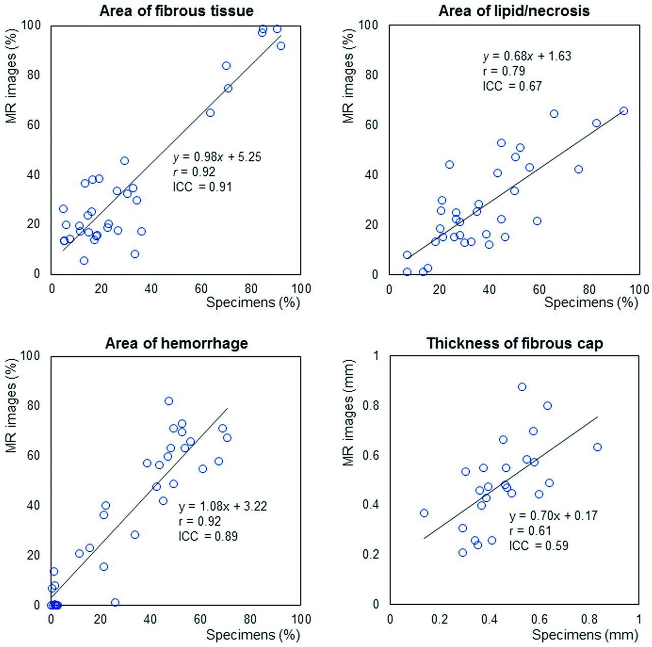
    Correlation and agreement between percentage area of intraplaque components and fibrous cap thickness observed in MR images and histologic specimens. Excellent positive correlation and agreement were observed between the percentage areas of the fibrous tissue and hemorrhagic areas within the plaque determined on MR images and histologic specimens. Percentage areas of lipid/necrosis and thickness of the fibrous cap also showed good correlation and agreement. r indicates the Pearson correlation coefficient.
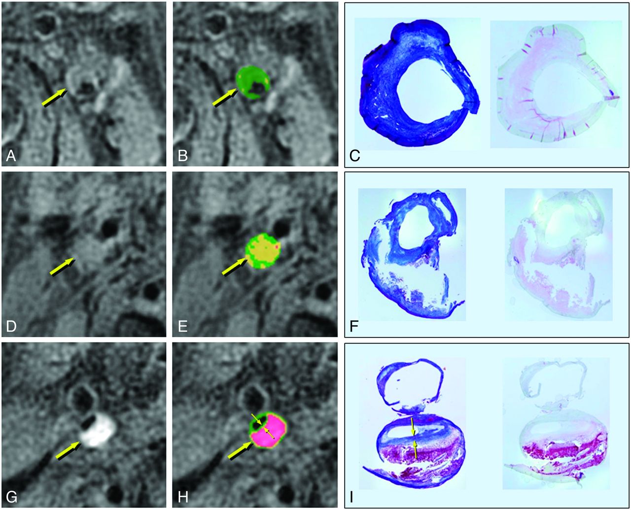
  • Fig 2.
    • Download figure
    • Open in new tab
    • Download powerpoint
    Fig 2.

    MR imaging and histologic findings of the carotid plaques. A, D, G, T1-weighted images. B, E, H, Color-coded maps. C, F, I, Corresponding histologic specimens (left, Masson trichrome staining; right, antiglycophorin-A staining). A–C, Left carotid stenosis in a 70-year-old man. The plaque shows isointensity to adjacent muscle on the T1-weighted image (A, arrow) and is mainly green on the color-coded map (B, arrow), suggesting a fibrous composition. On histologic examination, the corresponding plaque specimens consist mainly of thick fibrous tissue (C and D). The percentage areas of the fibrous, lipid/necrotic, and hemorrhagic components are 97%, 3%, and 0%, respectively, on MR images and 85%, 15%, and 0%, respectively, in the histologic specimens. D–F, Right carotid stenosis in a 68-year-old man. The plaque shows slight hyperintensity on the T1-weighted image (D, arrow) and is mainly yellow on the color map (E, arrow), suggesting a lipid-rich plaque. The corresponding plaque specimens contained lipid and necrotic tissue that flowed out of the specimen during the tissue preparation (F). The percentage areas of the fibrous, lipid/necrotic, and hemorrhagic components are 35%, 64%, and 1%, respectively, on the MR images, and 33%, 66%, and 1%, respectively, in the corresponding specimens. G–I, Right carotid stenosis in a 74-year-old man. The plaque shows marked hyperintensity on the T1-weighted image (G, arrow) and red on the color map (H, arrow), suggesting intraplaque hemorrhage. In the corresponding pathology specimens, the plaque contains massive hemorrhage with a thin fibrous cap (I). The percentage areas of fibrous, lipid/necrotic, and hemorrhagic components are 17%, 13%, and 70%, respectively, on MR imaging, and 15%, 33%, and 52%, respectively, in the specimens. The fibrous cap thicknesses on the MR images (H, small arrows) and in the specimen (I, small arrows) are 0.47 and 0.39 mm, respectively. (C,F, and I, 3x magnification.)

Tables

  • Figures
  • Percentage of areas of intraplaque components and fibrous cap thicknesses on color-coded MR images and histologic specimensa

    MR ImagesSpecimensrICC
    Fibrous tissue (%)5.7–98.7 (24.7)4.8–92.3 (21.0)0.920.91
    Lipid/necrosis (%)1.3–65.7 (22.2)7.0–93.8 (33.8)0.790.67
    Hemorrhage (%)0–82.0 (41.0)0–70.4 (35.8)0.920.89
    Fibrous cap (mm)0.21–0.87 (0.47)0.14–0.83 (0.45)0.610.59
    • Note:—r indicates the Pearson correlation coefficient.

    • ↵a Data are presented as range (median).

PreviousNext
Back to top

In this issue

American Journal of Neuroradiology: 35 (4)
American Journal of Neuroradiology
Vol. 35, Issue 4
1 Apr 2014
  • Table of Contents
  • Index by author
  • Complete Issue (PDF)
Advertisement
Print
Download PDF
Email Article

Thank you for your interest in spreading the word on American Journal of Neuroradiology.

NOTE: We only request your email address so that the person you are recommending the page to knows that you wanted them to see it, and that it is not junk mail. We do not capture any email address.

Enter multiple addresses on separate lines or separate them with commas.
Predicting Carotid Plaque Characteristics Using Quantitative Color-Coded T1-Weighted MR Plaque Imaging: Correlation with Carotid Endarterectomy Specimens
(Your Name) has sent you a message from American Journal of Neuroradiology
(Your Name) thought you would like to see the American Journal of Neuroradiology web site.
CAPTCHA
This question is for testing whether or not you are a human visitor and to prevent automated spam submissions.
Citation Tools
Predicting Carotid Plaque Characteristics Using Quantitative Color-Coded T1-Weighted MR Plaque Imaging: Correlation with Carotid Endarterectomy Specimens
S. Narumi, M. Sasaki, H. Ohba, K. Ogasawara, M. Kobayashi, T. Natori, J. Hitomi, H. Itagaki, T. Takahashi, Y. Terayama
American Journal of Neuroradiology Apr 2014, 35 (4) 766-771; DOI: 10.3174/ajnr.A3741

Citation Manager Formats

  • BibTeX
  • Bookends
  • EasyBib
  • EndNote (tagged)
  • EndNote 8 (xml)
  • Medlars
  • Mendeley
  • Papers
  • RefWorks Tagged
  • Ref Manager
  • RIS
  • Zotero
Share
Predicting Carotid Plaque Characteristics Using Quantitative Color-Coded T1-Weighted MR Plaque Imaging: Correlation with Carotid Endarterectomy Specimens
S. Narumi, M. Sasaki, H. Ohba, K. Ogasawara, M. Kobayashi, T. Natori, J. Hitomi, H. Itagaki, T. Takahashi, Y. Terayama
American Journal of Neuroradiology Apr 2014, 35 (4) 766-771; DOI: 10.3174/ajnr.A3741
del.icio.us logo Digg logo Reddit logo Twitter logo CiteULike logo Facebook logo Google logo Mendeley logo
  • Tweet Widget
  • Facebook Like
  • Google Plus One
Purchase

Jump to section

  • Article
    • Abstract
    • ABBREVIATIONS:
    • Materials and Methods
    • Results
    • Discussion
    • Conclusions
    • Footnotes
    • References
  • Figures & Data
  • Info & Metrics
  • References
  • PDF

Related Articles

  • No related articles found.
  • PubMed
  • Google Scholar

Cited By...

  • Optimal MR Plaque Imaging for Cervical Carotid Artery Stenosis in Predicting the Development of Microembolic Signals during Exposure of Carotid Arteries in Endarterectomy: Comparison of 4 T1-Weighted Imaging Techniques
  • Imaging the Intracranial Atherosclerotic Vessel Wall Using 7T MRI: Initial Comparison with Histopathology
  • Carotid Plaque Characterization Using 3D T1-Weighted MR Imaging with Histopathologic Validation: A Comparison with 2D Technique
  • Crossref
  • Google Scholar

This article has not yet been cited by articles in journals that are participating in Crossref Cited-by Linking.

More in this TOC Section

  • Association of Left Vertebral Artery Hypoplasia with Posterior Circulation Stroke and the Functional Outcome of Patients with Atrial Fibrillation–Related Cardioembolic Stroke
  • MRI Detection of Carotid Intraplaque Hemorrhage and Postintervention Cognition
  • Survey of the American Society of Neuroradiology Membership on the Use and Value of Extracranial Carotid Vessel Wall MRI
Show more Extracranial Vascular

Similar Articles

Advertisement

News and Updates

  • Lucien Levy Best Research Article Award
  • Thanks to our 2022 Distinguished Reviewers
  • Press Releases

Resources

  • Evidence-Based Medicine Level Guide
  • How to Participate in a Tweet Chat
  • AJNR Podcast Archive
  • Ideas for Publicizing Your Research
  • Librarian Resources
  • Terms and Conditions

Opportunities

  • Share Your Art in Perspectives
  • Get Peer Review Credit from Publons
  • Moderate a Tweet Chat

American Society of Neuroradiology

  • Neurographics
  • ASNR Annual Meeting
  • Fellowship Portal
  • Position Statements

© 2023 by the American Society of Neuroradiology | Print ISSN: 0195-6108 Online ISSN: 1936-959X

Powered by HighWire