Advertisement

Main menu

  • Home
  • Content
    • Current Issue
    • Publication Preview--Ahead of Print
    • Past Issue Archive
    • Case of the Week Archive
    • Classic Case Archive
    • Case of the Month Archive
    • COVID-19 Content and Resources
  • For Authors
  • About Us
    • About AJNR
    • Editors
    • American Society of Neuroradiology
  • Submit a Manuscript
  • Podcasts
    • Subscribe on iTunes
    • Subscribe on Stitcher
  • More
    • Subscribers
    • Permissions
    • Advertisers
    • Alerts
    • Feedback
  • Other Publications
    • ajnr

User menu

  • Subscribe
  • Alerts
  • Log in

Search

  • Advanced search
American Journal of Neuroradiology

American Journal of Neuroradiology

  • Subscribe
  • Alerts
  • Log in

Advanced Search

  • Home
  • Content
    • Current Issue
    • Publication Preview--Ahead of Print
    • Past Issue Archive
    • Case of the Week Archive
    • Classic Case Archive
    • Case of the Month Archive
    • COVID-19 Content and Resources
  • For Authors
  • About Us
    • About AJNR
    • Editors
    • American Society of Neuroradiology
  • Submit a Manuscript
  • Podcasts
    • Subscribe on iTunes
    • Subscribe on Stitcher
  • More
    • Subscribers
    • Permissions
    • Advertisers
    • Alerts
    • Feedback
  • Follow AJNR on Twitter
  • Visit AJNR on Facebook
  • Follow AJNR on Instagram
  • Join AJNR on LinkedIn
  • RSS Feeds
Research ArticlePediatrics

Diffusion Imaging for Tumor Grading of Supratentorial Brain Tumors in the First Year of Life

S.F. Kralik, A. Taha, A.P. Kamer, J.S. Cardinal, T.A. Seltman and C.Y. Ho
American Journal of Neuroradiology April 2014, 35 (4) 815-823; DOI: https://doi.org/10.3174/ajnr.A3757
S.F. Kralik
aFrom Indiana University School of Medicine, Department of Radiology and Imaging Sciences, Indianapolis, Indiana.
  • Find this author on Google Scholar
  • Find this author on PubMed
  • Search for this author on this site
A. Taha
aFrom Indiana University School of Medicine, Department of Radiology and Imaging Sciences, Indianapolis, Indiana.
  • Find this author on Google Scholar
  • Find this author on PubMed
  • Search for this author on this site
A.P. Kamer
aFrom Indiana University School of Medicine, Department of Radiology and Imaging Sciences, Indianapolis, Indiana.
  • Find this author on Google Scholar
  • Find this author on PubMed
  • Search for this author on this site
J.S. Cardinal
aFrom Indiana University School of Medicine, Department of Radiology and Imaging Sciences, Indianapolis, Indiana.
  • Find this author on Google Scholar
  • Find this author on PubMed
  • Search for this author on this site
T.A. Seltman
aFrom Indiana University School of Medicine, Department of Radiology and Imaging Sciences, Indianapolis, Indiana.
  • Find this author on Google Scholar
  • Find this author on PubMed
  • Search for this author on this site
C.Y. Ho
aFrom Indiana University School of Medicine, Department of Radiology and Imaging Sciences, Indianapolis, Indiana.
  • Find this author on Google Scholar
  • Find this author on PubMed
  • Search for this author on this site
  • Article
  • Figures & Data
  • Info & Metrics
  • References
  • PDF
Loading

Article Figures & Data

Figures

  • Tables
  • Fig 1.
    • Download figure
    • Open in new tab
    • Download powerpoint
    Fig 1.

    A 4-month-old girl with poorly differentiated carcinoma of the left cerebral hemisphere. A, Representative axial image from ADC map demonstrates manual tracing with general exclusion of large areas of blood products represented by susceptibility artifacts. B, Histogram of all included ADC values of the tumor from the semi-automated method.

  • Fig 2.
    • Download figure
    • Open in new tab
    • Download powerpoint
    Fig 2.

    An 11-month-old boy with atypical teratoid/rhabdoid tumor. A, Axial T2-weighted image demonstrates a heterogeneous mass in the right frontal temporal lobe with peripheral cystic change, little peritumoral white matter T2 prolongation, and B, heterogeneous enhancement on postcontrast axial 3D T1-weighted image. C, Representative ADC manual region of interest measurement with a small region of interest within the lowest signal portion of the tumor and larger region of interest measuring the contralateral normal thalamus. D, Semi-automated histogram for the ADC values of the entire tumor.

  • Fig 3.
    • Download figure
    • Open in new tab
    • Download powerpoint
    Fig 3.

    A 12-month-old child with desmoplastic infantile ganglioglioma. A, Axial T2-weighted image demonstrates a heterogeneous mass in the left frontal lobe with cystic change, adjacent T2 prolongation, midline shift, and contralateral ventricular entrapment. B, Axial 3D T1-weighted image demonstrates a peripheral enhancing solid component along the dural margin. C, ADC map with representative manual region of interest evaluation within the solid components of the tumor and contralateral white matter. D, Semi-automated histogram for the ADC values of the entire tumor.
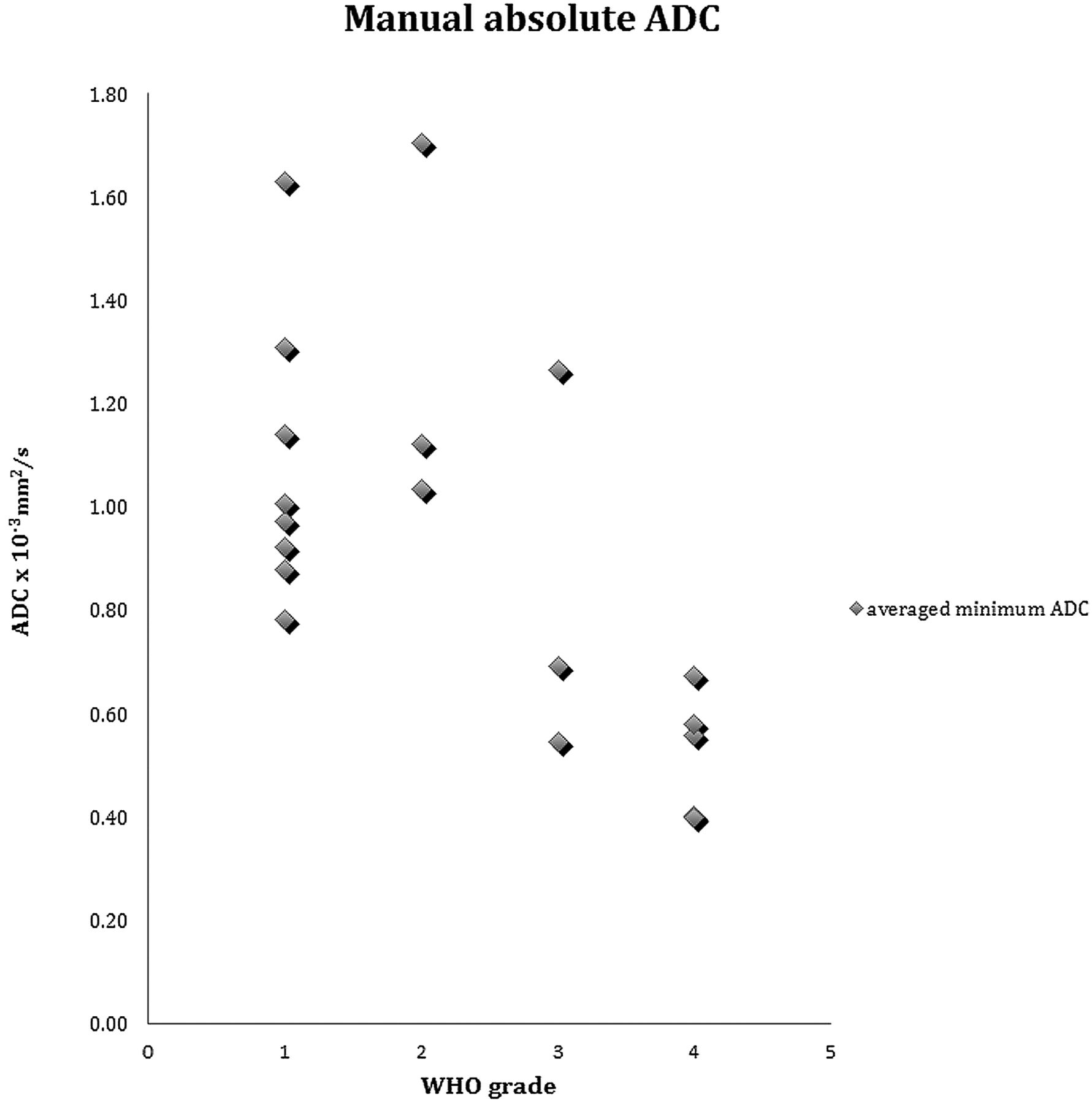
  • Fig 4.
    • Download figure
    • Open in new tab
    • Download powerpoint
    Fig 4.

    Scatterplot of the average absolute minimum ADC for all tumors by WHO grading.

  • Fig 5.
    • Download figure
    • Open in new tab
    • Download powerpoint
    Fig 5.

    Receiver operating characteristic curve for manual absolute ADC demonstrates a significant area above the 50% diagonal. Threshold according to the Youden index is ≤0.698 × 10⁻³ mm2/s for WHO grade III and IV tumors versus grade I and II tumors.

  • Fig 6.
    • Download figure
    • Open in new tab
    • Download powerpoint
    Fig 6.

    A, Summation semi-automated histograms of ADC values of all low-grade tumors compared with high-grade tumors. B, Overlay histograms of all low-grade tumors. C, Overlay histograms of all high-grade tumors.

Tables

  • Figures
    • View popup
    Table 1:

    Tumor pathology with corresponding WHO grade and ADC values from manual ROI measurement

    Tumor TypeWHO GradeAverage Minimum ADC × 10−3 mm2/sADC Ratio ThalamusADC Ratio White Matter
    Desmoplastic infantile gangliogliomaI1.631.681.50
    Desmoplastic infantile gangliogliomaI0.881.050.79
    Desmoplastic infantile gangliogliomaI0.971.080.80
    Desmoplastic infantile gangliogliomaI1.140.870.76
    Choroid plexus papillomaI1.011.051.10
    Choroid plexus papillomaI0.921.160.79
    Choroid plexus papillomaI1.311.531.23
    Pilocytic astrocytomaI0.780.960.75
    Astrocytoma, focalII1.711.501.35
    Pilomyxoid astrocytomaII1.031.060.71
    Astrocytoma, diffuseII1.121.270.98
    Anaplastic ependymomaIII0.690.820.59
    Anaplastic ependymoma with tanycytic featuresIII0.540.570.47
    Choroid plexus carcinomaIII1.270.930.90
    Atypical teratoid/rhabdoid tumorIV0.670.530.41
    Atypical teratoid/rhabdoid tumorIV0.400.550.42
    Poorly differentiated carcinomaIV0.560.450.32
    Poorly differentiated carcinomaIV0.400.510.35
    GlioblastomaIV0.580.620.41
    P value, t test between low- and high-grade groups.0018<.0001.00042
    • Note:—All 3 parameters were significant between the difference of the means of the low-grade and high-grade groups. Of note, the choroid plexus carcinoma in our study is an outlier in the high-grade group.

    • View popup
    Table 2:

    Semi-automated metrics including mean and standard deviations for low- and high-grade tumors

    Low-GradeHigh-GradeP Value (t test)
    Mean (×10−3 mm2/s)Standard Deviation (×10−3 mm2/s)Mean (×10−3 mm2/s)Standard Deviation (×10−3 mm2/s)
    Average1.7170.3771.4590.341.138
    Standard deviation0.4580.2470.6160.189.135
    Skew0.8030.7531.3720.704.111
    Kurtosis1.1292.5151.9802.754.501
    Peak location1.5180.4931.0750.275.023a
    Peak height, normalized0.0880.0410.0690.029.258
    5th Percentile1.130.1510.7780.267.007a
    10th Percentile1.180.1510.8580.260.008a
    25th Percentile1.320.1801.0160.278.018a
    50th Percentile1.680.4531.2700.349.037a
    75th Percentile2.010.5981.7920.564.411
    90th Percentile2.320.6812.3630.591.903
    95th Percentile2.500.7052.6960.541.518
    95th to 5th Percentile1.3770.7041.9180.560.080
    50th to 5th Percentile0.5550.4250.4910.273.695
    95th to 50th Percentile0.8210.4341.4260.362.004a
    • ↵a Significant P value.

PreviousNext
Back to top

In this issue

American Journal of Neuroradiology: 35 (4)
American Journal of Neuroradiology
Vol. 35, Issue 4
1 Apr 2014
  • Table of Contents
  • Index by author
  • Complete Issue (PDF)
Advertisement
Print
Download PDF
Email Article

Thank you for your interest in spreading the word on American Journal of Neuroradiology.

NOTE: We only request your email address so that the person you are recommending the page to knows that you wanted them to see it, and that it is not junk mail. We do not capture any email address.

Enter multiple addresses on separate lines or separate them with commas.
Diffusion Imaging for Tumor Grading of Supratentorial Brain Tumors in the First Year of Life
(Your Name) has sent you a message from American Journal of Neuroradiology
(Your Name) thought you would like to see the American Journal of Neuroradiology web site.
CAPTCHA
This question is for testing whether or not you are a human visitor and to prevent automated spam submissions.
Citation Tools
Diffusion Imaging for Tumor Grading of Supratentorial Brain Tumors in the First Year of Life
S.F. Kralik, A. Taha, A.P. Kamer, J.S. Cardinal, T.A. Seltman, C.Y. Ho
American Journal of Neuroradiology Apr 2014, 35 (4) 815-823; DOI: 10.3174/ajnr.A3757

Citation Manager Formats

  • BibTeX
  • Bookends
  • EasyBib
  • EndNote (tagged)
  • EndNote 8 (xml)
  • Medlars
  • Mendeley
  • Papers
  • RefWorks Tagged
  • Ref Manager
  • RIS
  • Zotero
Share
Diffusion Imaging for Tumor Grading of Supratentorial Brain Tumors in the First Year of Life
S.F. Kralik, A. Taha, A.P. Kamer, J.S. Cardinal, T.A. Seltman, C.Y. Ho
American Journal of Neuroradiology Apr 2014, 35 (4) 815-823; DOI: 10.3174/ajnr.A3757
del.icio.us logo Digg logo Reddit logo Twitter logo CiteULike logo Facebook logo Google logo Mendeley logo
  • Tweet Widget
  • Facebook Like
  • Google Plus One
Purchase

Jump to section

  • Article
    • Abstract
    • ABBREVIATION:
    • Materials and Methods
    • Results
    • Discussion
    • Conclusions
    • Footnotes
    • References
  • Figures & Data
  • Info & Metrics
  • References
  • PDF

Related Articles

  • No related articles found.
  • PubMed
  • Google Scholar

Cited By...

  • Grading of Pediatric Intracranial Tumors: Are Intravoxel Incoherent Motion and Diffusional Kurtosis Imaging Superior to Conventional DWI?
  • Neuroimaging Appearance of Cerebral Malignant Epithelioid Glioneuronal Tumors in Children
  • Contrast Leakage Patterns from Dynamic Susceptibility Contrast Perfusion MRI in the Grading of Primary Pediatric Brain Tumors
  • Crossref
  • Google Scholar

This article has not yet been cited by articles in journals that are participating in Crossref Cited-by Linking.

More in this TOC Section

  • Common Neuroimaging Findings in Bosch-Boonstra-Schaaf Optic Atrophy Syndrome
  • Longitudinal MRI Evaluation of Brain Development in Fetuses with Congenital Diaphragmatic Hernia around the Time of Fetal Endotracheal Occlusion
  • Early Fetal Corpus Callosum: Demonstrating Normal Growth and Detecting Pathologies in Early Pregnancy
Show more Pediatrics

Similar Articles

Advertisement

News and Updates

  • Lucien Levy Best Research Article Award
  • Thanks to our 2022 Distinguished Reviewers
  • Press Releases

Resources

  • Evidence-Based Medicine Level Guide
  • How to Participate in a Tweet Chat
  • AJNR Podcast Archive
  • Ideas for Publicizing Your Research
  • Librarian Resources
  • Terms and Conditions

Opportunities

  • Share Your Art in Perspectives
  • Get Peer Review Credit from Publons
  • Moderate a Tweet Chat

American Society of Neuroradiology

  • Neurographics
  • ASNR Annual Meeting
  • Fellowship Portal
  • Position Statements

© 2023 by the American Society of Neuroradiology | Print ISSN: 0195-6108 Online ISSN: 1936-959X

Powered by HighWire